3D human induced pluripotent stem cell–derived bioengineered skeletal muscles for tissue, disease and therapy modeling
Luca Pinton, Moustafa Khedr, Valentina M. Lionello, Shilpita Sarcar, Sara M. Maffioletti, Sumitava Dastidar, Elisa Negroni, SungWoo Choi, Noreen Khokhar, Anne Bigot, John R. Counsell, Andreia Sofia Bernardo, Peter S. Zammit, Francesco Saverio Tedesco
induced pluripotent stem cells
bioengineered skeletal muscles
3D hydrogels
muscle pathology
therapy modeling


Supplementary information
Supplementary Information
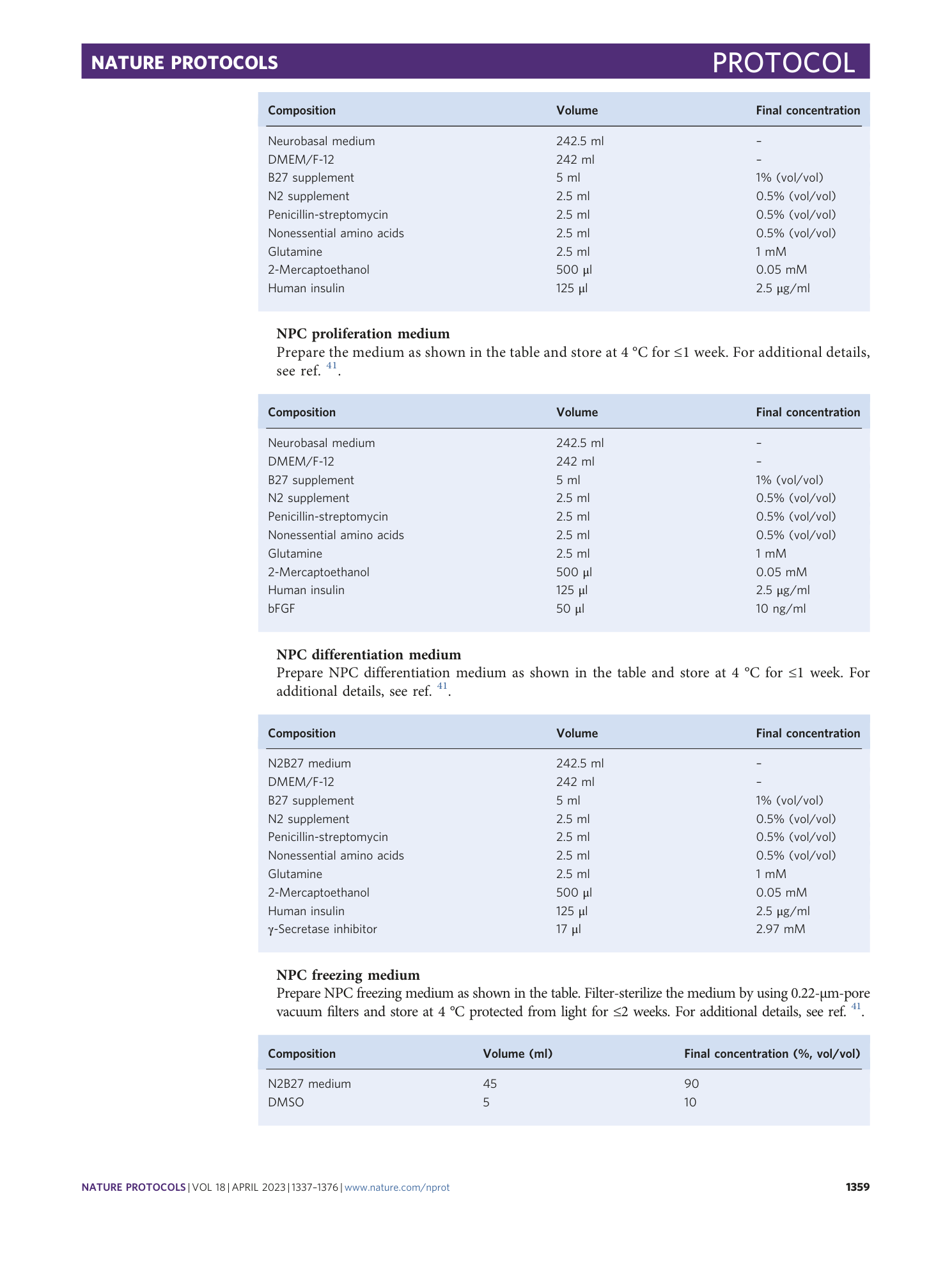
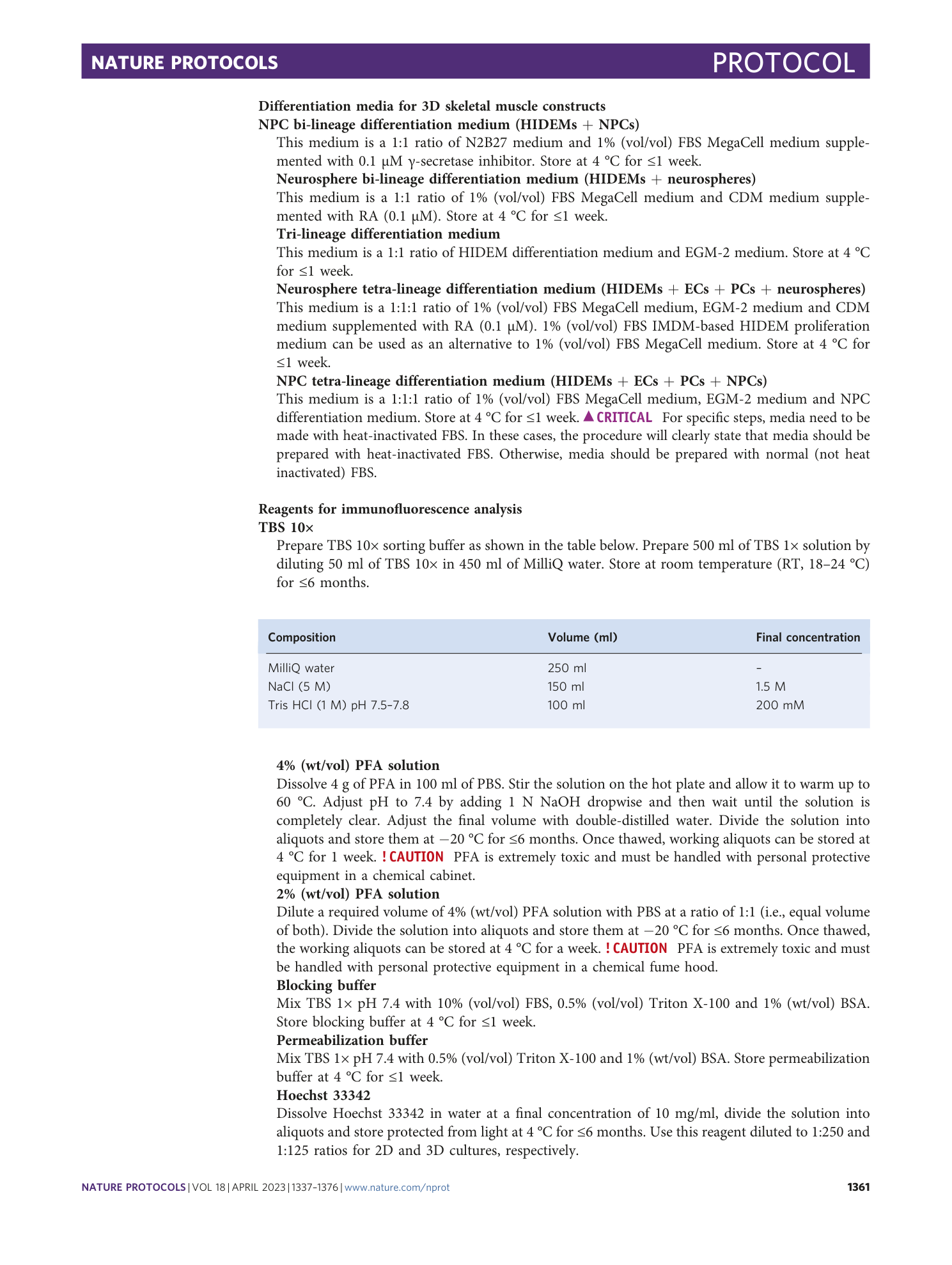
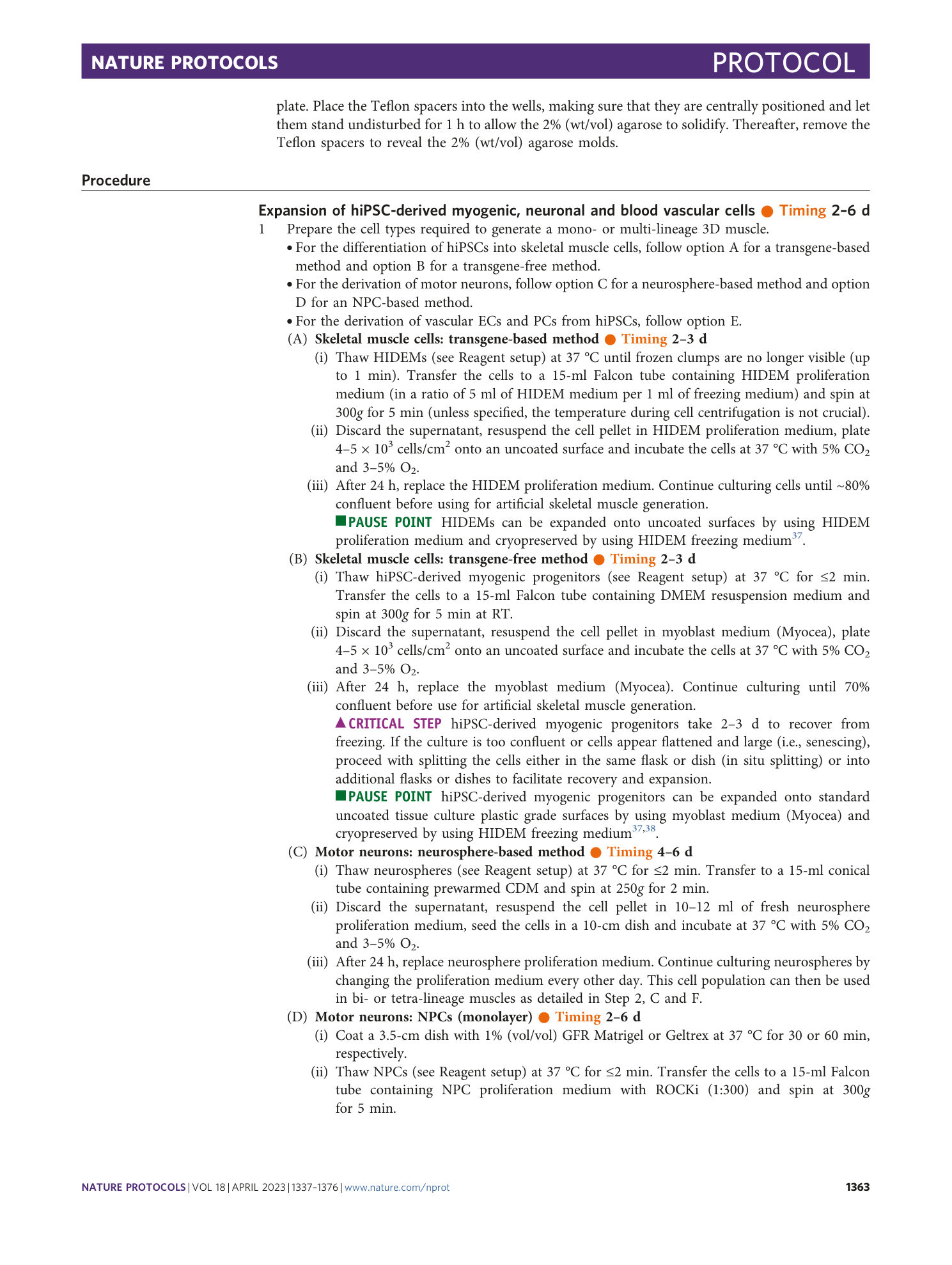
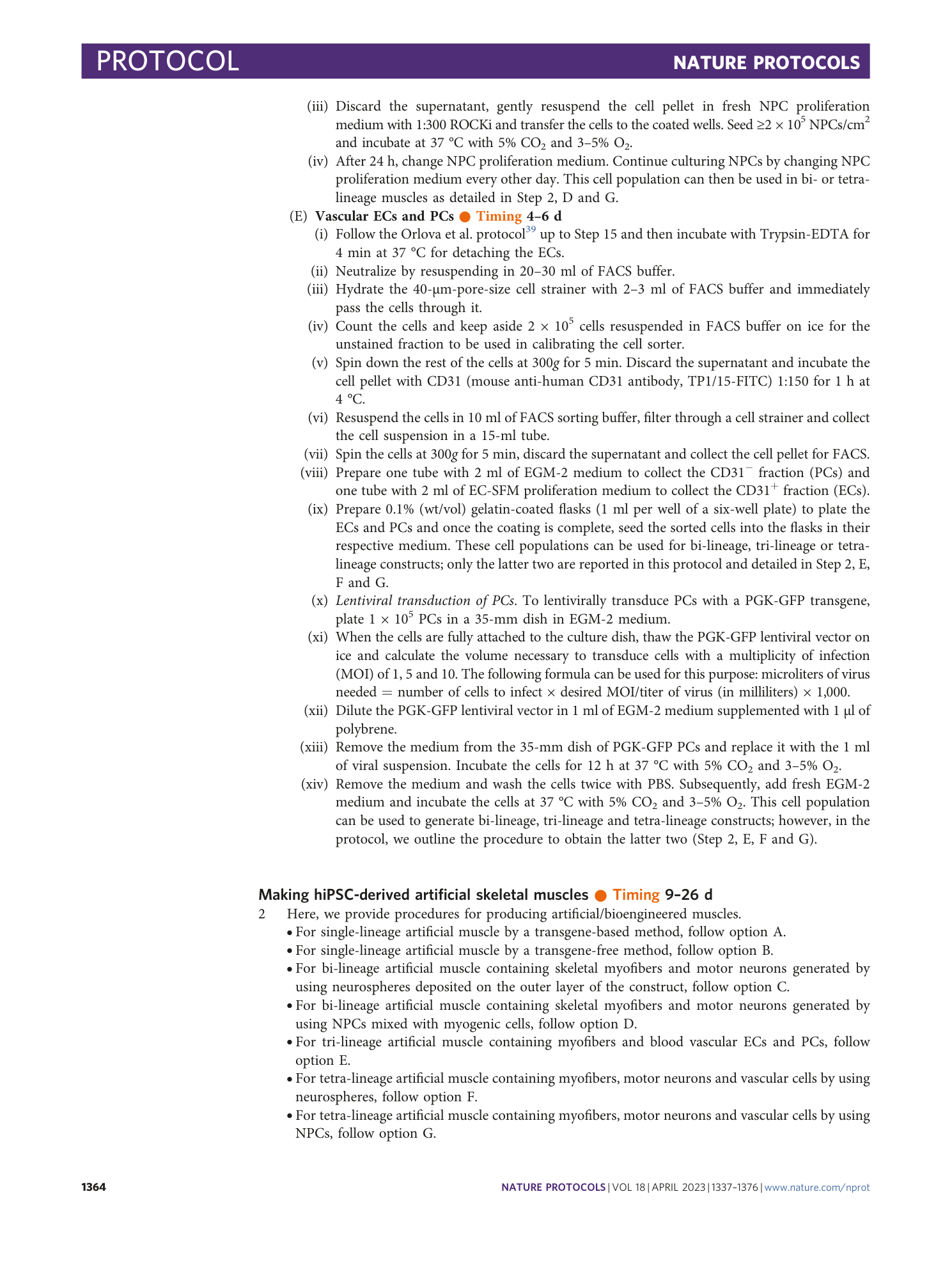
Supplementary Table 1 and Protocol 1
Supplementary Video 1
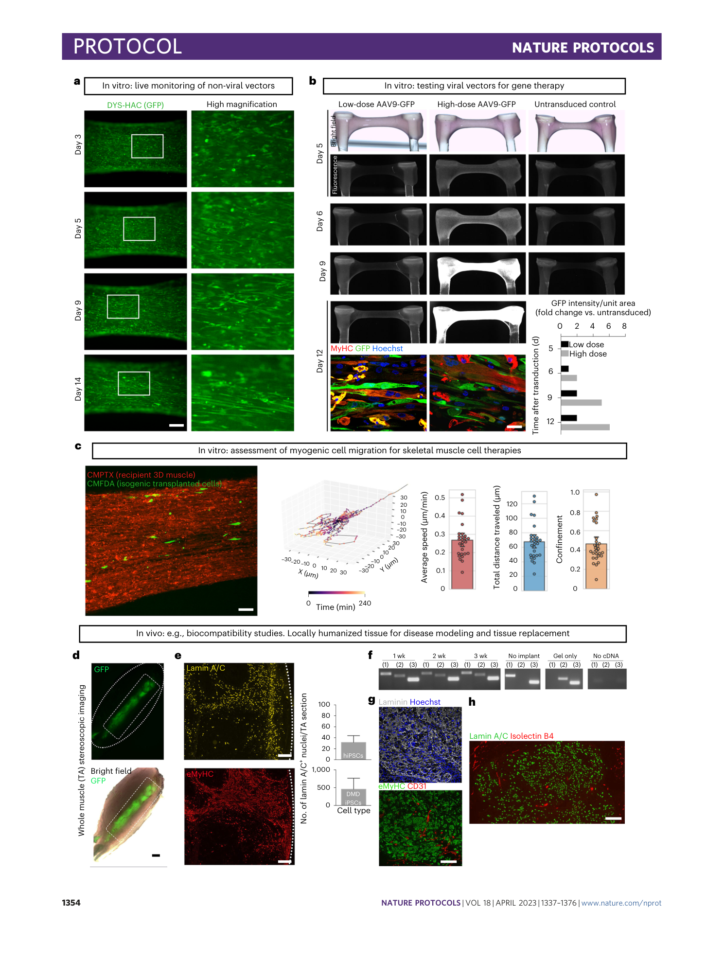
Real-time video of DMD hiPSC-derived 3D skeletal muscle construct contractions in response to 10-V electrical stimulation at 1 Hz
Supplementary Video 2
Calcium transients in DMD hiPSC-derived 3D artificial muscles incubated with the fluorescence Ca 2+ indicator Fluo-4AM upon electrical stimulation. During the first 10 s, no electrical stimuli were applied to the muscle. At second 10, the 3D muscle was stimulated with 10 V at 0.33 Hz, and at second 20, the 3D muscle was stimulated with 20 V at 0.33 Hz
Supplementary Video 3
Maximum-intensity projection time-lapse video of 5-chloromethylfluorescein diacetate (CMFDA)-stained HIDEMs (green) deposited on hiPSC-derived 3D muscles (red). 100-µm stacks of muscles were imaged every 12 min for 4 h by using a spinning disk microscope. Scale bar, 100 µm
Supplementary Video 4
3D reconstruction of a 3D artificial muscle imaged with a light-sheet microscope immunostained as detailed in the advanced imaging section of Fig. 2
Supplementary Video 5
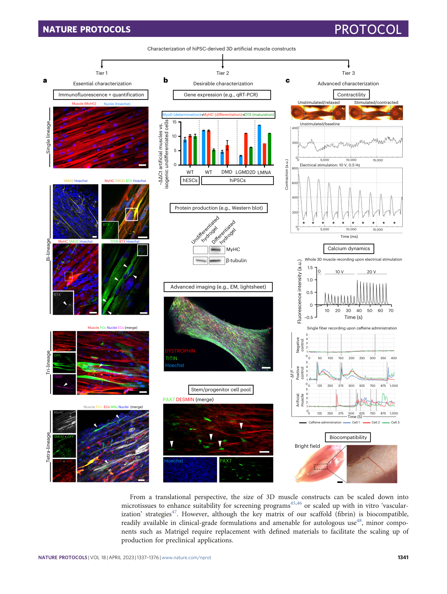
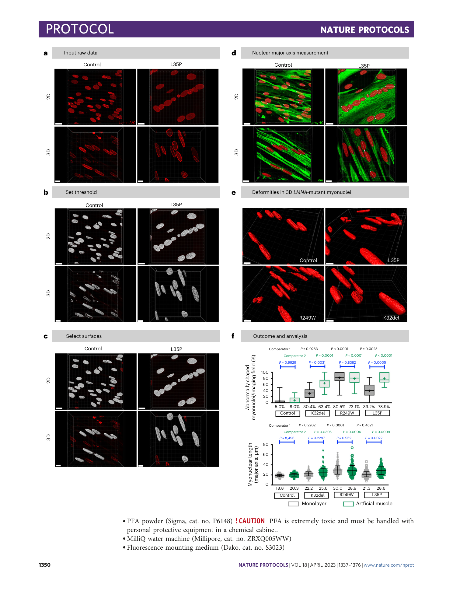
3D reconstruction nuclear morphology (lamin A/C in red) and myotubes (eMyHC in green) of a healthy control hiPSC-derived 3D muscle construct shown in Fig. 3. The 3D reconstruction was done by using Imaris software and confocal z-stacks of immunostained artificial muscles. Video reproduced from ref. 29 under Creative Commons license CC BY 4.0
Supplementary Video 6
3D reconstruction nuclear morphology (lamin A/C in red) and myotubes (eMyHC in green) of an LMNA-mutant (R249W) hiPSC-derived 3D muscle construct shown in Fig. 3. The 3D reconstruction was done by using Imaris software and confocal z-stacks of immunostained artificial muscles. Video reproduced from ref. 29 under Creative Commons license CC BY 4.0
