Spatial analysis of tissue immunity and vascularity by light sheet fluorescence microscopy
Duo Zhang, Abigail H. Cleveland, Elisavet Krimitza, Katherine Han, Chenlong Yi, Andrea L. Stout, Wei Zou, Jay F. Dorsey, Yanqing Gong, Yi Fan


Extended
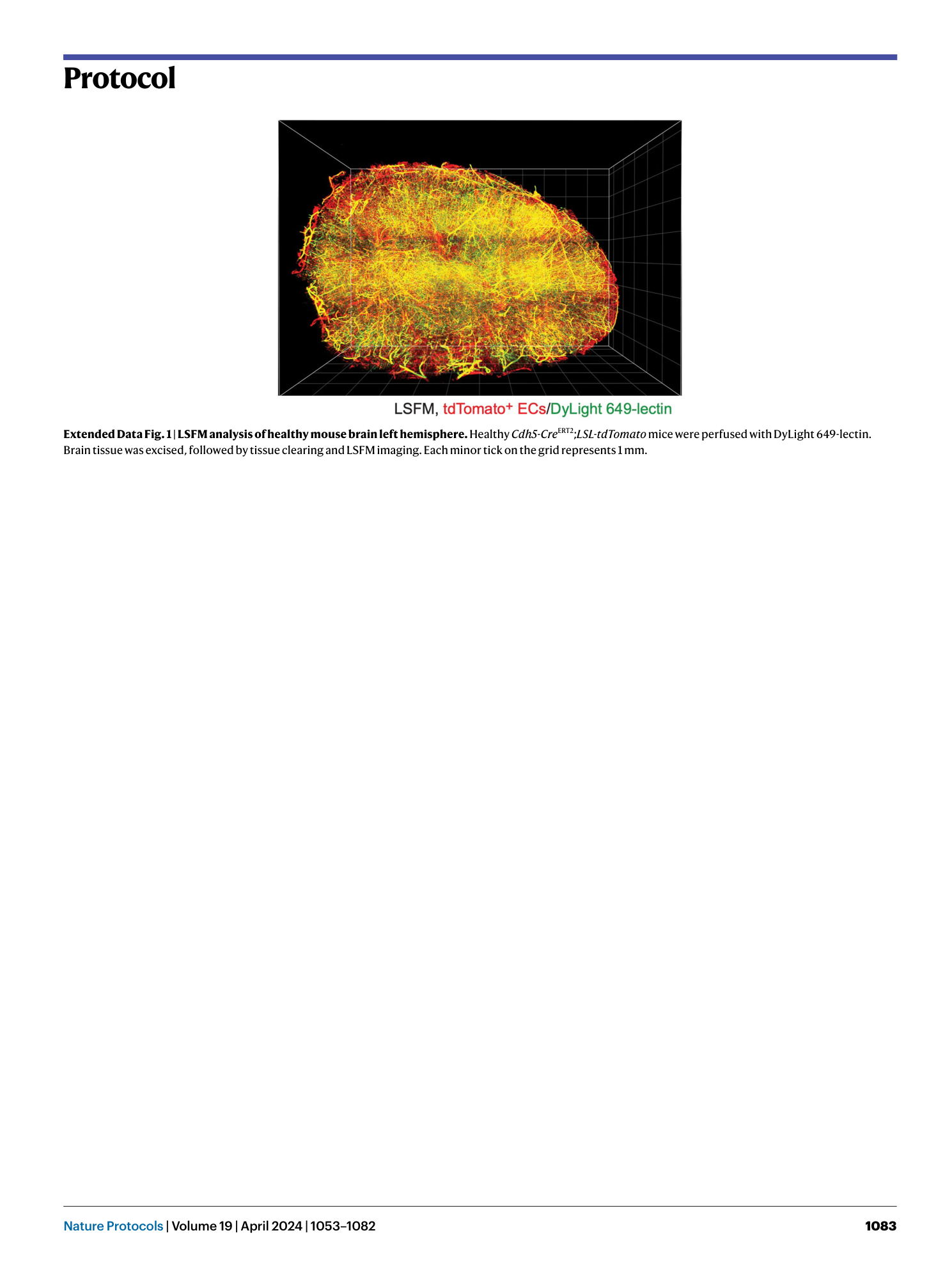
Extended Data Fig. 1 LSFM analysis of healthy mouse brain left hemisphere.
Healthy Cdh5-Cre ERT2 ; LSL-tdTomato mice were perfused with DyLight 649-lectin. Brain tissue was excised, followed by tissue clearing and LSFM imaging. Each minor tick on the grid represents 1 mm.
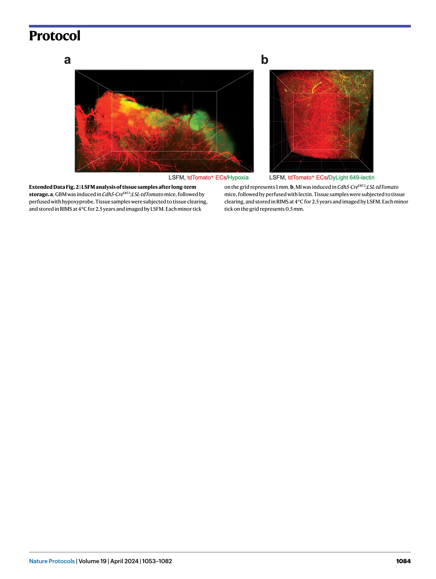
Extended Data Fig. 2 LSFM analysis of tissue samples after long-term storage.
a , GBM was induced in Cdh5-Cre ERT2 ; LSL-tdTomato mice, followed by perfused with hypoxyprobe. Tissue samples were subjected to tissue clearing, and stored in RIMS at 4°C for 2.5 years and imaged by LSFM. Each minor tick on the grid represents 1 mm. b , MI was induced in Cdh5-Cre ERT2 ; LSL-tdTomato mice, followed by perfused with lectin. Tissue samples were subjected to tissue clearing, and stored in RIMS at 4°C for 2.5 years and imaged by LSFM. Each minor tick on the grid represents 0.5 mm.
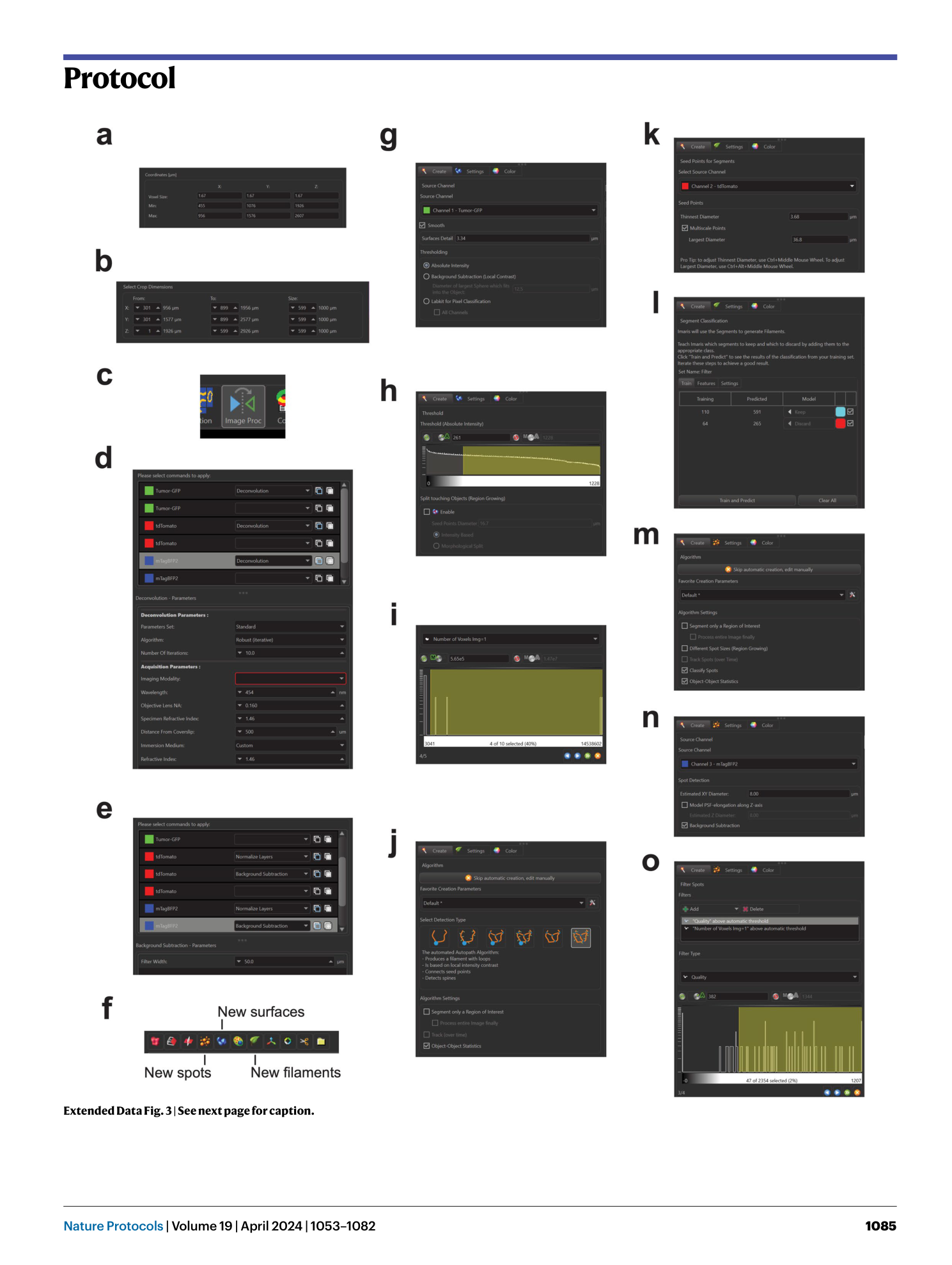
Extended Data Fig. 3 Key steps of image processing and analysis in Imaris.
a , Voxel size correction in “Image Properties” (Step 42). b , Crop out ROI in “Crop 3D” (Step 43). c , “Image Processing” module (Step 44). d , Setting deconvolution parameters in the “Image Processing” module (Step 44). e , Preprocessing the image using “Normalize Layers” and “Background Subtraction “ (Steps 45, 46). f , Creating objects for feature registration (Step 48). g , Initial parameters for creating surface objects (Step 48A(ii)). h , Thresholding of surface object creation (Step 48A(iii)). i , Filtering generated surface objects by size (Step 48A(iv)). j , Multiscale Points method of generating filament seed points (Step 48B(ii)). k , Seed point filtering by vessel diameter (Step 48B(ii)). l , Segment classification and filtering by machine learning-based model (Step 48B(iii)). m , Initial parameters for creating spot objects (Step 48C(i)). n , Spot detection by feature diameter (Step 48C(ii)). o , Filter Spot feature by quality and diameter/size (Step 48C(iii–iv)).
Supplementary information
Reporting Summary
Supplementary Software 1
3D printing file for ETC sample holder.
Supplementary Software 2
3D printing file for sample imaging holder.
