Mixotrophy - Quantification of the percent of phytoplankton cells with preys (e.g. fluorescent microspheres or fluorescently labeled bacteria)
Valeria Jimenez
Abstract
This methodology describes a protocol modified from Sherr and Sherr (1993) to determine phytoplankton prey uptake using flow cytometry and yellow-green fluorescent polystyrene-based microspheres (YG-beads; 0.5 µm in diameter, Fluoresbrite, Polysciences, Inc., Warrington, PA, USA) or fluorescently labelled bacteria (FLBs) as food proxies. FLBs were prepared according to the protocol of Sherr et al . (1987) using the bacteria Brevundimonas diminuta (strain CECT313, also named Pseudomonas diminuta ), obtained from the Spanish Type Culture Collection (CECT, Valencia, Spain).
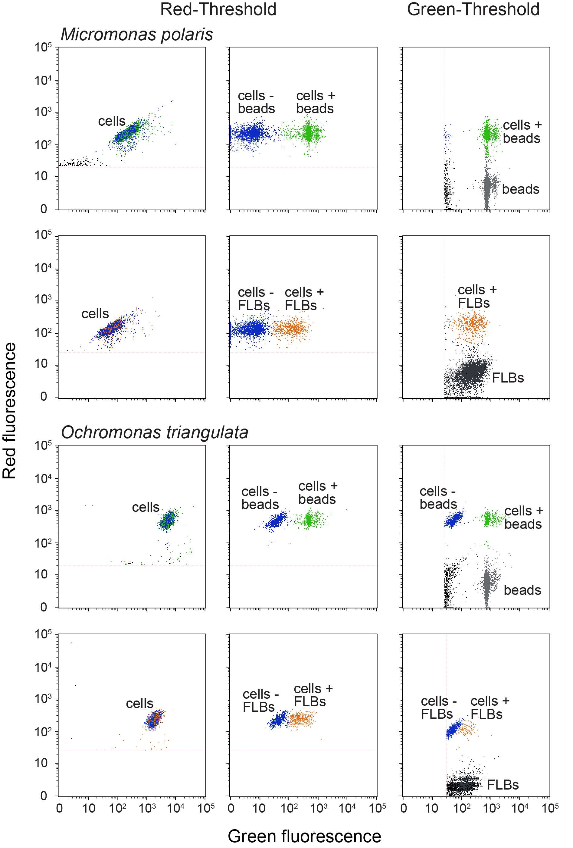
The percent of cells with prey were measured in the laboratory in four Micromonas polaris strains and one mixotrophic strain ( Ochromonas triangulata ) used as a positive control. The percent of cells with beads was determined in samples fixed with Lugol's iodine solution (protocol explained in detail here) and enumerated with a flow cytometer (Guava easyCyte, Luminex Corporation, USA) where cells that contained chlorophyll as well as green fluorescence (same signal as the prey added, YG-beads or FLBs) were considered to be cells containing prey (quantification protocol explain in detail here). To determine the uptake of prey over time the percent of cells with preys was quantified in each experimental flask by first adding preys and then sub-sampling and fixing after an incubation of 0 (T0), 20 (T20) and 40 (T40) minutes. The T0 sample accounts for the physical attachment of preys to the cell and therefore the percent of cells ingesting prey corresponds to the percent of cells with prey at T20 or T40, minus the percent of cells with prey at T0.
References References:
Sherr, EB. & Sherr, BF. (1993). Protistan grazing rates via uptake of fluorescently labeled prey. In Kemp, P., Sherr, B., Sherr, E. & Cole, J. (eds.) Handbook of Methods in Aquatic Microbial Ecology, 695–701 (Lewis Publishers: Boca Raton, USA).
Sherr, EB., Sherr, BF. & Fallon, RD. (1987). Use of monodispersed, fluorescently labeled bacteria to estimate in situ protozoan bacterivory. Appl Environ Microbiol.
Before start
Steps
Experiment Set up and Fixation
Prepare your prey (YG-beads or FLBs) stock solution. Vortex vigorously and sonicate the solution for ~60 seconds.
Take the phytoplankton flasks under different culture conditions (e.g. phytoplankton incubated in the dark and under nutrient limitation) on which to test uptake of prey (use at least 2 to 3 replicates per phytoplankton strain and culture condition)
Add the preys to each flask (one flask at a time). After microsphere addition, gently mix the flask to evenly distribute microspheres and then take a sub-sample from the flask for immediate fixation (see fixation protocol section). This sub-sample is the T0 for background correction (attachment of preys to cells not associated with mixotrophy)
After the addition of preys and the sampling and fixation of the T0 sample, each flask with preys can be sampled and fixed over time (After 20 (T20) and 40 (T40) minutes incubation with preys) as performed with T0 sample.
After all flasks and time points are fixed, samples are kept on ice are in the dark to be run in the flow cytometer the same day.
Quantification of the percent of cells with preys using a flow cytometer
After all flasks and time points are fixed, samples are run in the flow cytometer the same day.
To determine the percent of cells with preys the sample is run with the threshold on red fluorescence. Cells without preys only show red autofluorescence from chlorophyll and the cells with the same chlorophyll fluorescence as well as green fluorescence (same signal as the prey added, YG-beads or FLBs) are considered to be cells containing prey.
In addition, to confirm the total concentration of prey added to each flask, the sample was also run with the threshold on green fluorescence

Fixation protocol: Acid Lugol’s iodine solution
The following fixation protocol must be done under a hood.
The volume of reagents must be added to 5 ml of culture.
Add 50 µl of acid Lugol’s iodine solution (see fixatives preparation section) to the sample.
Add 300 µl of 37% Formaldehyde to the sample.
Add 50 µl 3% Thiosulfate (see fixatives preparation section) to the sample.
Make sure the sample is thoroughly cleared of Lugol’s stain, if not add more sodium thiosulfate until it is clear (e.g. another 50 µl)
Fixatives preparation: acid Lugol's solution
Dissolve 10 g of Potassium iodide in 100 ml distilled water
Slowly add 5 g of iodine crystals. Shake to dissolve.
Add 10 ml glacial acetic acid. Store in the dark.
Fixatives preparation: Sodium Thiosulfate at 3% weight/volume
Add 3 ml of sodium thiosulfate in 97 ml of distilled water

