SEM-EDS Protocol V1
Diana Vanegas, Saylen Sofia Paz, David Bahamon-Pinzon
Abstract
This protocol outlines the methodology for conducting Scanning Electron Microscopy (SEM) and Energy-Dispersive X-ray Spectroscopy (EDS) analysis of laser-inscribed graphene (LIG) electrodes using a SU5000 microscope. The procedure details the preparation of LIG electrodes, their imaging under SEM, and subsequent elemental analysis via EDS. By combining SEM imaging for morphology assessment with EDS for elemental composition identification, this protocol facilitates a comprehensive characterization of LIG electrodes, crucial for understanding their structural features and properties in various applications. The fusion of SEM and EDS techniques offers insights into the intricate structure-property relationships of LIG electrodes, advancing their potential in diverse fields such as energy storage, sensing, and electronics.
Attachments
Steps
Overview
Protocol SEM/EDS 1.1 Scanning Electron Microscopy (SEM) and Energy-Dispersive X-ray Spectroscopy (EDS) analysis Saylen Sofia Paz, David Bahamon-Pinzon, Diana Vanegas Gamboa Clemson University This protocol describes the process of performing the Scanning Electron Microscopy (SEM) and Energy-Dispersive X-ray Spectroscopy (EDS) analysis of laser-inscribed graphene (LIG) electrodes (e.g. Protocol L-Cu1.1) using a Scanning Electron Microscope SU5000. Fig 1 depicts the process for the analysis. The time must be change according with the number of the samples that you have.

MATERIALS
- LIG sensors (Protocol L-Cu1.1)
- Tweezers
- Tape. NEM. Nisshin em.co.ltd
- Sampleholder
- USB
HARDWARE
- Scanning Electron Microscope SU5000.
Procedure
Step 1) Turn on the computer and equipment screens
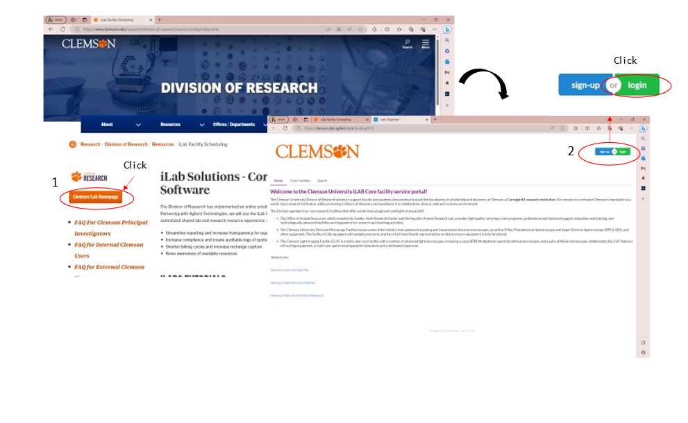
- Login from any device with internet access to iLab's website (iLabFacilityScheduling (clemson.edu))
- Log into your account
- Click on the start button Note: Automatically, the screens of the equipment will turn on (Figure 2)
Step 2) Preparation of the sample
- Take a piece of carbon tape and put it in a 51 mm holder
- Remove the layer that is covering the tape
- Place the samples over the tape using the tweezers (Fig 3A)
- Put the bottom part of the holder (Fig 3B)
- Verify that the height of the holder and the sample is within the range of the scale (Fig 3C) Note: Try to organize the samples with something that you can recognize each sample
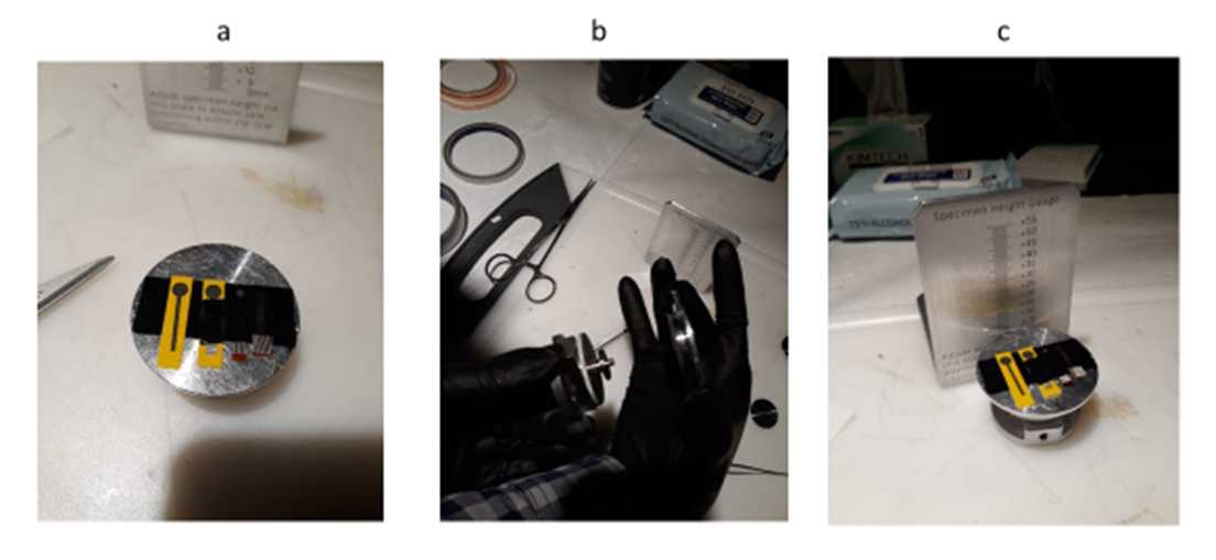
Step 3) Set up the equipment
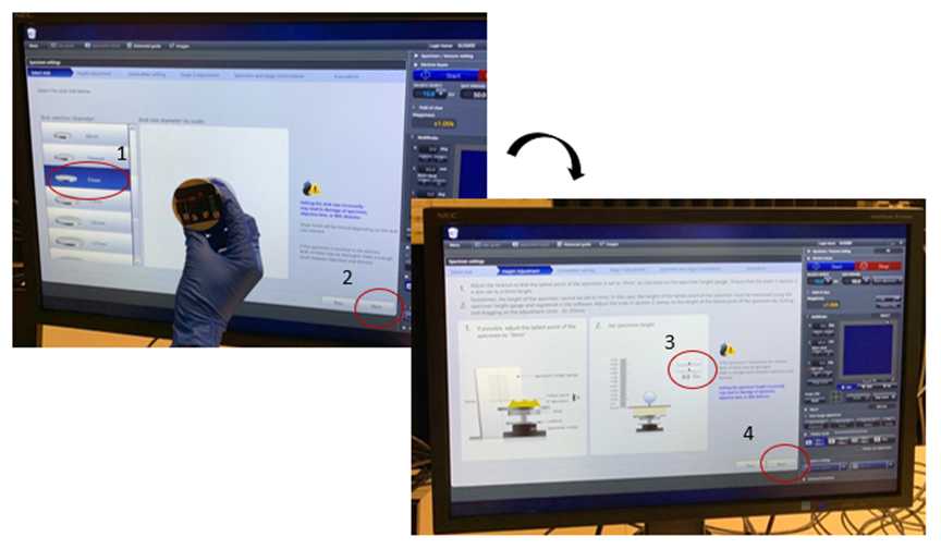
- The equipment is always under high vacuum. Letting air in the equipment is necessary to open the chamber and place the sample. To do this, click Air in the Specimen/vacuum setting (Figure 4)
- The equipment should start beeping. Wait for the process to be completed. It beeps three times
- Click Specimen settings in the popup window (See Figure 4)
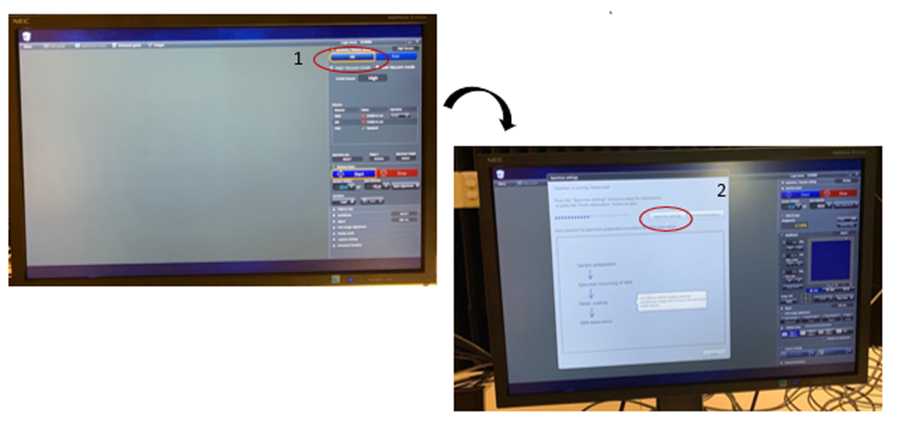
3.2 CameraNavi setting (optional)
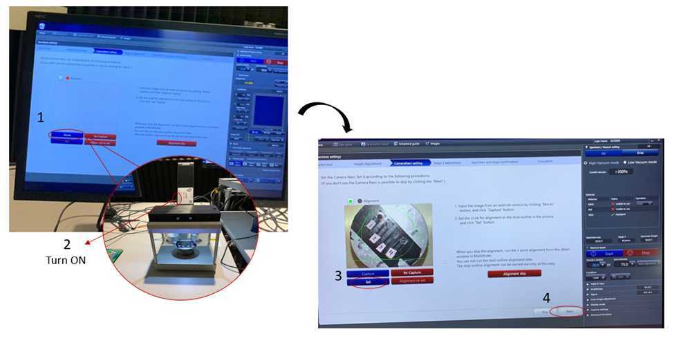
- Turn the light of the camera on with the switch
- Put the holder on the camera. The holder must reach the top (Fig 3)
- Click movie
- Push the buttons to zoom in or out
- Click capture
- The holder should fit the circle in the screen. Move with the mouse.
- The triangle and circle in the screen should fit the holder
- Click set
- Green should show up
- Click next (Figure 6)
3.3 Placeholder in the chamber
-
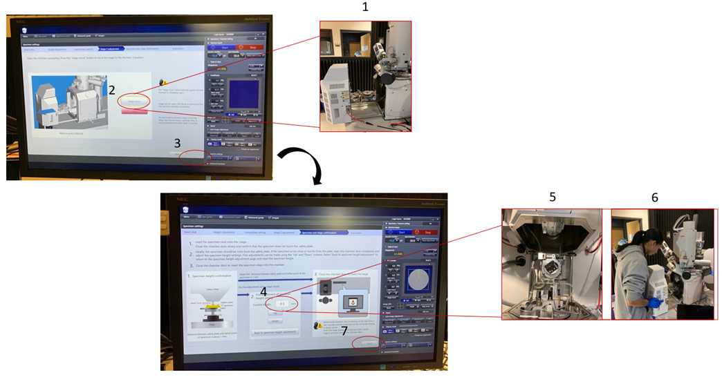
Pull the gate of the chamber all the way out until you hear a switch (Fig 7)
-
Click stage move Note: It starts beeping as it moves up the stage
-
When it stops beeping, put a sample. The holder must reach the top. The top is gate located next to the wall of the room
-
Close the chamber slowly and verify the height of the holder. The sample should be under the plate
-
If the sample is too tall, pull the equipment out again until you hear the click and click down as needed.
-
Close the door entirely until hearing a slight beep.
-
Click Next (See Figure 7)
Step 5) Insert detector
- Verify that Detector BSE-All is selected in the top right
- Check that BSE can load in the field specimen
- BSE (Back-scattered Electron Detector) able to load
- Insert the detector by turning slowly in the “IN” direction until it stops (Figure 10)
- Click OK in the pop-up window
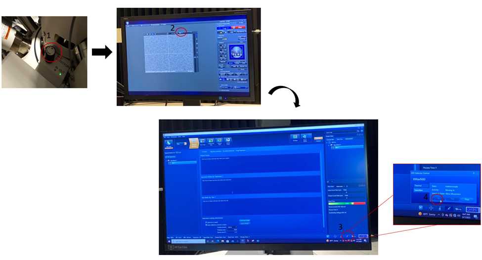
Step 6)Calibrate the beam (brightness, contrast, focus)
Note: There are several parameters that can be adjusted from the box of the equipment
-
Brightturn knob inc
-
Contrast
-
Speeds o 1. Fast, 2. Slow, 3. Red (reduced). Click 2 twice to change to 2.1.
-
Focus
-
Coarse
-
Stigma(stism)
-
Click mode to change to alignment
-
Loo for something to focus
-
When finding something, click 1. Bum aligment
-
Center beam by adjusting X and Y knobs
-
If nothing happens when clicking auto bright, check alignment
-
Click mode
-
Set an appropriate picture for alignment. The focus should not move after this
-
Aperture alignment
-
Moving alignment
-
Move x. Then it doesn’t move.
-
Click mode
-
Stigma alignment for X
-
Click mode. I f it looks fine, push mode again. Note: Always focus at high magnification (e.g. more than 25X)
-
VP: low vacuum (vacuum pressure)
-
60 Pa
-
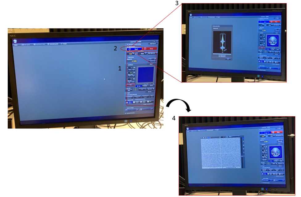
Click start
Step 7) Set capture settings
-
Go to capture settings in the bottom right corner of the screen
-
Save settings Select the folder "Vanegas" Note: If is necessary, create the folder
-
Click to capture settings
-
In capture settings, select: o Speed: slow
o Image size: 1280x960 (adequate for papers, not
for posters or magazines. For posters, use 5120x3840)
o Time: 16 s. This corresponds approximately to Slow 3
Step 8) Take picture
- In the box, press Capt to capture pictures. Note: After each capture, the software changes to Freeze. Press 4 in the box of commands to return to Run.
Step 9) Set settings for EDS
- Change voltage to 15 kV. 5 kV is proper for carbon but not for platinum or copper. Note: The working distance should be 10 mm. For example, If the distance in the screen is 6.2 mm, we need 4 mm more. We need to add 4 mm to the value of Z (in
Specimen/Vacuum settings). If Z was 5 at the beginning, set a Z of 9.
- 7.4-10=2.5
Step 10) Open the software for EDS
-
In the second screen, open the software Aztec. Open project LIG (create a new folder or select the folder “Vanegas”)
-
Process time: 6. Note: The process time of 1 is the slowest, and 6 is the highest.
-
Element distribution
Step 11) Final parameters and start
- Verify that the dead time is around 40%
- Select EDS-SEM in the top left area of the screen.
- Set Analyze: overall.
- Select Point 81D (scan specific position). Other options include color map, line graph
- Click New specimen
- Click New sample
- Uncheck box Specimen is coated
- Click Scan image
- Click Settings
- Select a Scan size of 1024
- Dwell t (time/pixel): 5 μs
- In the “sun” at the bottom right corner, the brightness can be adjusted
- Click Start
Step 12) Remove the sample from the chamber
- When the analysis is completed, click “Stop”
- Take the detector out by tunning the knob in the “OUT” direction until it stops.
- In Vacuum, select Air.
- Click Finish
- Pull the gate of the chamber to open the equipment.
- Take the sample holder out.
- Close the chamber.
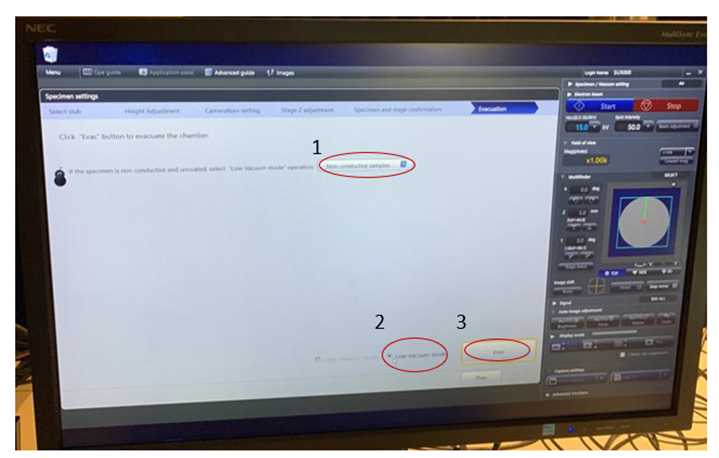
- Click “Evac” (the equipment must remain in High vacuum when not used).
- Clean the working station
Notes
"dark"areas have lower average atomic number* Detectors: o BSE is used to find differences in composition. High energy elements.
o Secondary electrons (SE) has lower energy. It is used to study the sample surface and texture (this detector didn’t work well because of the substrate for LIG samples)
- A higher pressure from a low vacuum setting will scatter the electrons, and the image will be distorted
- For EDS, 5 kV is adequate for Carbon but not for copper or platinum
- For elements such copper, platinum and gold an adequate potential ranges 15 – 20 kV
- For LIG samples, it is adequate to work in non-conductive samples and low vacuum
- For high resolution images, it is recommended to use the autofocus and auto contrast from the program and slow 3
