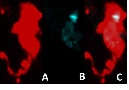
Immunostaining of Bodo saltans
Ewa Chrostek, Mastaneh Ahrar, Gregory Dd Hurst
Abstract
This protocol is used in our Laboratory in Liverpool to perform IF on Bodo saltans cells.
Steps
Culture conditions
Bodo saltans was cultured in a cerophyl-based medium enriched with 3.5 mM sodium phosphate dibasic (Na2HPO4)1. Cultures were incubated at 22 °C in T25 tissue culture flasks containing 20 ml of media bacterized with Klebsiella pneumoniae subsp. Pneumoniae (ATCC® 700831).
Immunostaining
Please, follow the steps below to prepare cells for immunostaining.
Filter the culture through 100 and 8 µm filter.
Incubate with secondary antibody (1:1000, diluted in PTX + 5% FBS) overnight at 4°C.
Rinse and wash 3 times for 1 hour with PTX at room temperature. During 2nd wash add Hoechest 33342 (Thermo Fisher, 1:2000) for 10 minutes. This will be rinsed away with the 3rd wash.
Remove the agarose from the well with clean forceps and place it on a microscope slide.
Add a drop of a mounting medium (eg. Vectashield, Vector Laboratories), and flatten the agarose as much as you can using the coverslip.
Proceed with either fluorescence or confocal imaging.

Harvest the cells by centrifugation at 1200 × g for 12 mins at 19 °C.
Wash the cells with PBS and centrifuge as described above.
Dissolve the pellet in 15 µl of PBS and mix with the same volume of low melting temperature agarose (eg. Thermo Fisher Scientific) in a single well of a 96-well plate. Let it ser for a few seconds.
Add 200 µl of 4% PFA and incubate at room temperature for 10 minutes or at 4 °C overnight.
Wash with PTX (PBS + 0,1% TritonX) 4 times, 30 mins each wash.
Block in 5% FBS+PTX overnight at 4 °C.
Incubate with primary antibody (1:100, diluted in PTX + 5% FBS), for 5 hours at room temperature or overnight at 4 °C.
Rinse and wash 3 times for 1 hour with PTX at room temperature.
References
Gomaa Fatma, Li ZuHong, Docampo Roberto, Girguis Peter, E. V. Bodo saltans culture protocol V.2. Protocols.io (2018). doi:10.17504/protocols.io.sh6eb9e


