Generation, functional analysis and applications of isogenic three-dimensional self-aggregating cardiac microtissues from human pluripotent stem cells
Giulia Campostrini, Viviana Meraviglia, Elisa Giacomelli, Ruben W. J. van Helden, Loukia Yiangou, Richard P. Davis, Milena Bellin, Valeria V. Orlova, Christine L. Mummery
isogenic microtissues
human pluripotent stem cells
cardiac differentiation
3D tissue models
disease modeling


Supplementary information
Supplementary Information
Supplementary Figs. 1–3.
Reporting Summary
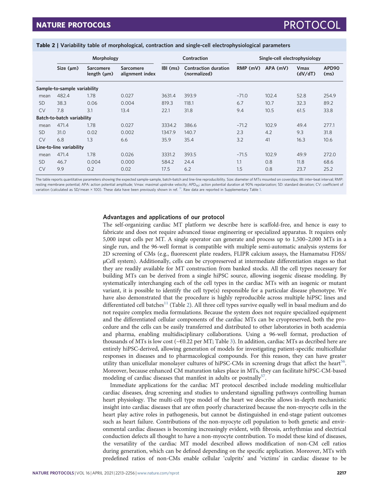
Supplementary Table 1
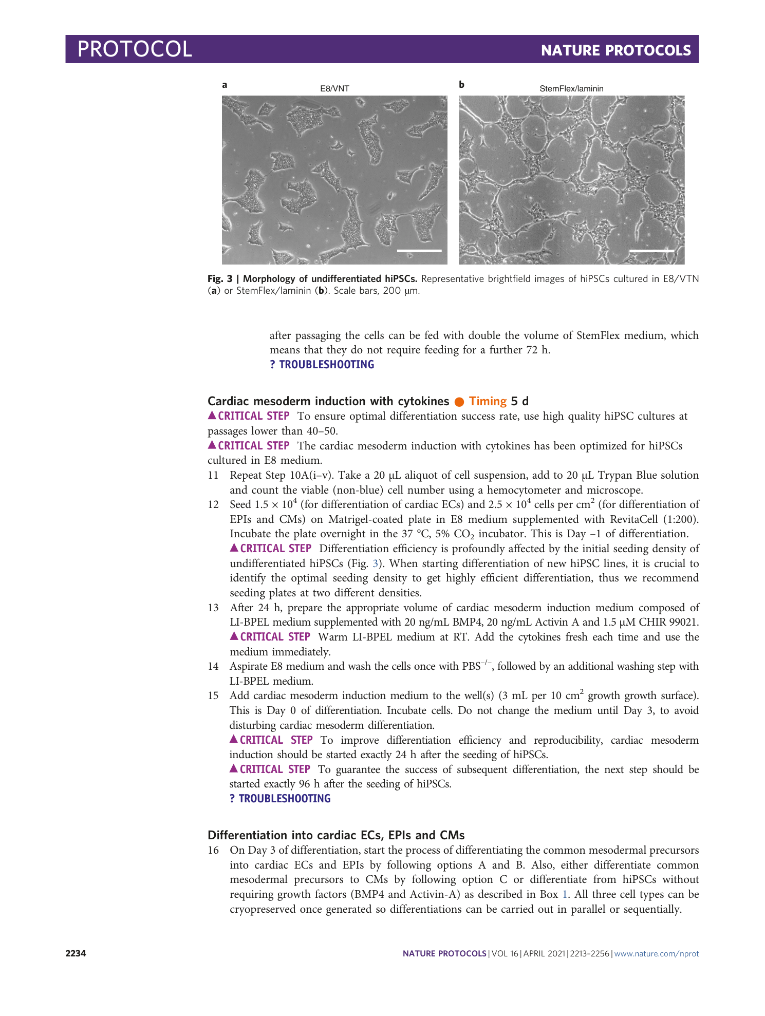
Raw data for results summarized in Table 3. Table contains morphological, contraction and single-cell electrophysiological parameters measured in LUMC0099iCTRL04 and LUMC0020iCTRL06 hiPSC lines.
Supplementary Video 1
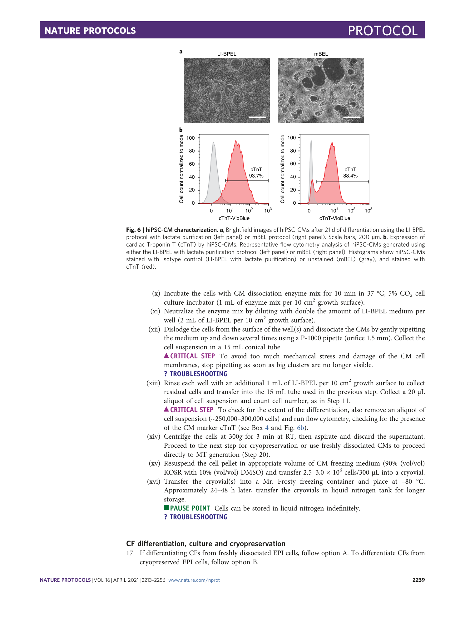
Beating monolayer hiPSC-CMs after 21 d of differentiation using LI-BPEL protocol with lactate purification
Supplementary Video 2
Beating monolayer hiPSC-CMs after 21 d of differentiation using mBPEL protocol
Supplementary Video 3
Time-lapse movie of the first 72 h of MT formation (time frame: 15 min)
Supplementary Video 4
Movie of representative MT showing CD31 positive ECs organized in a vessel-like structure (in red) and CMs stained with ACTN2 (in green) (15 f.p.s.)
Supplementary Video 5
Contracting MT paced at 1 Hz at after 21 d in culture
Supplementary Video 6
Contracting MT loaded with Fluo-4, paced at 1.5 Hz after 21 d in culture
