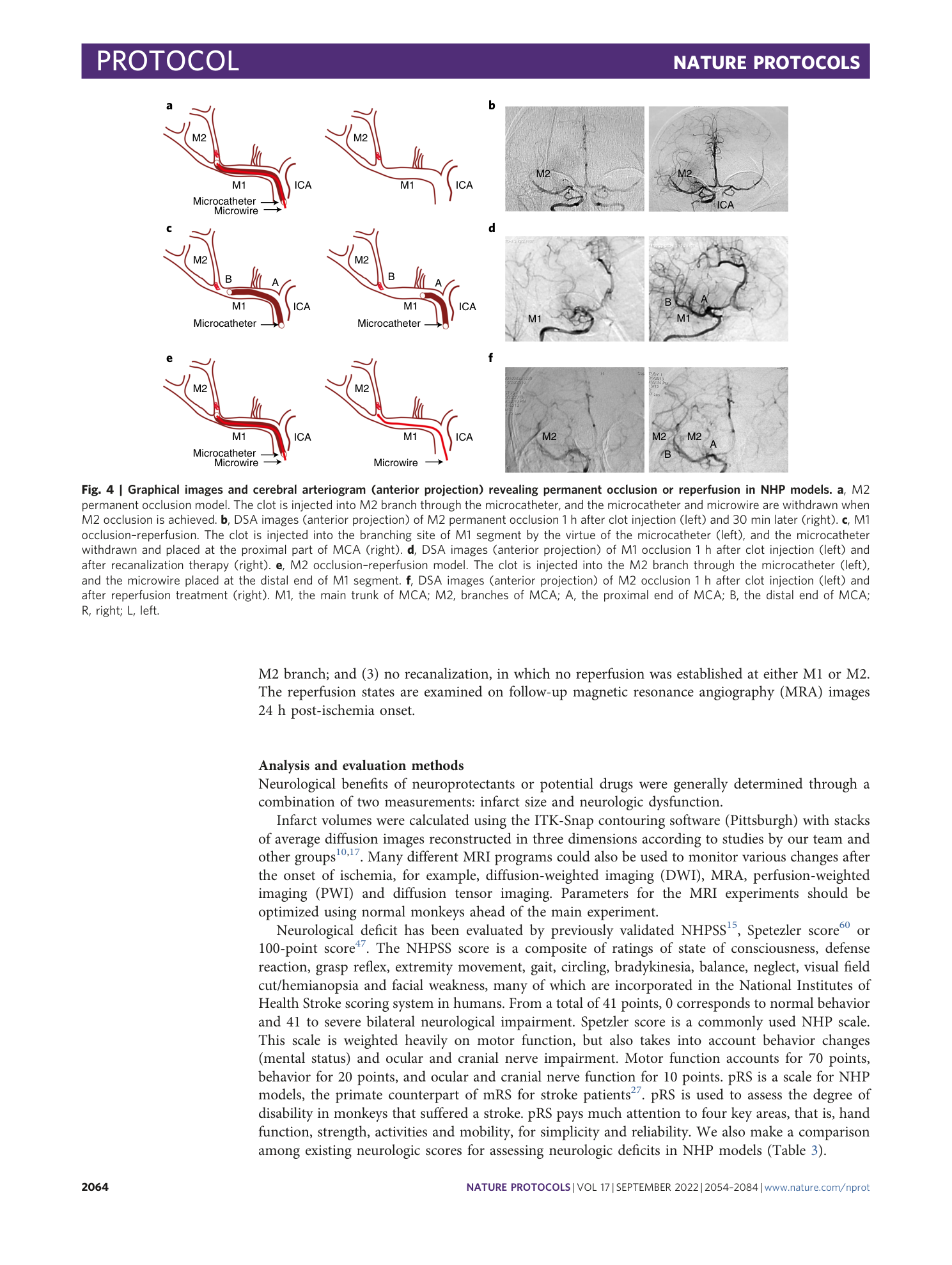
A clinically relevant model of focal embolic cerebral ischemia by thrombus and thrombolysis in rhesus monkeys
Di Wu, Jian Chen, Longfei Wu, Hangil Lee, Jingfei Shi, Mo Zhang, Yanhui Ma, Xiaoduo He, Zixin Zhu, Feng Yan, Chuanjie Wu, Yunxia Duan, Yongjuan Fu, Sijie Li, Xinglong Zhi, Xuxiang Zhang, Shengli Li, Yuchuan Ding, Xunming Ji


Extended
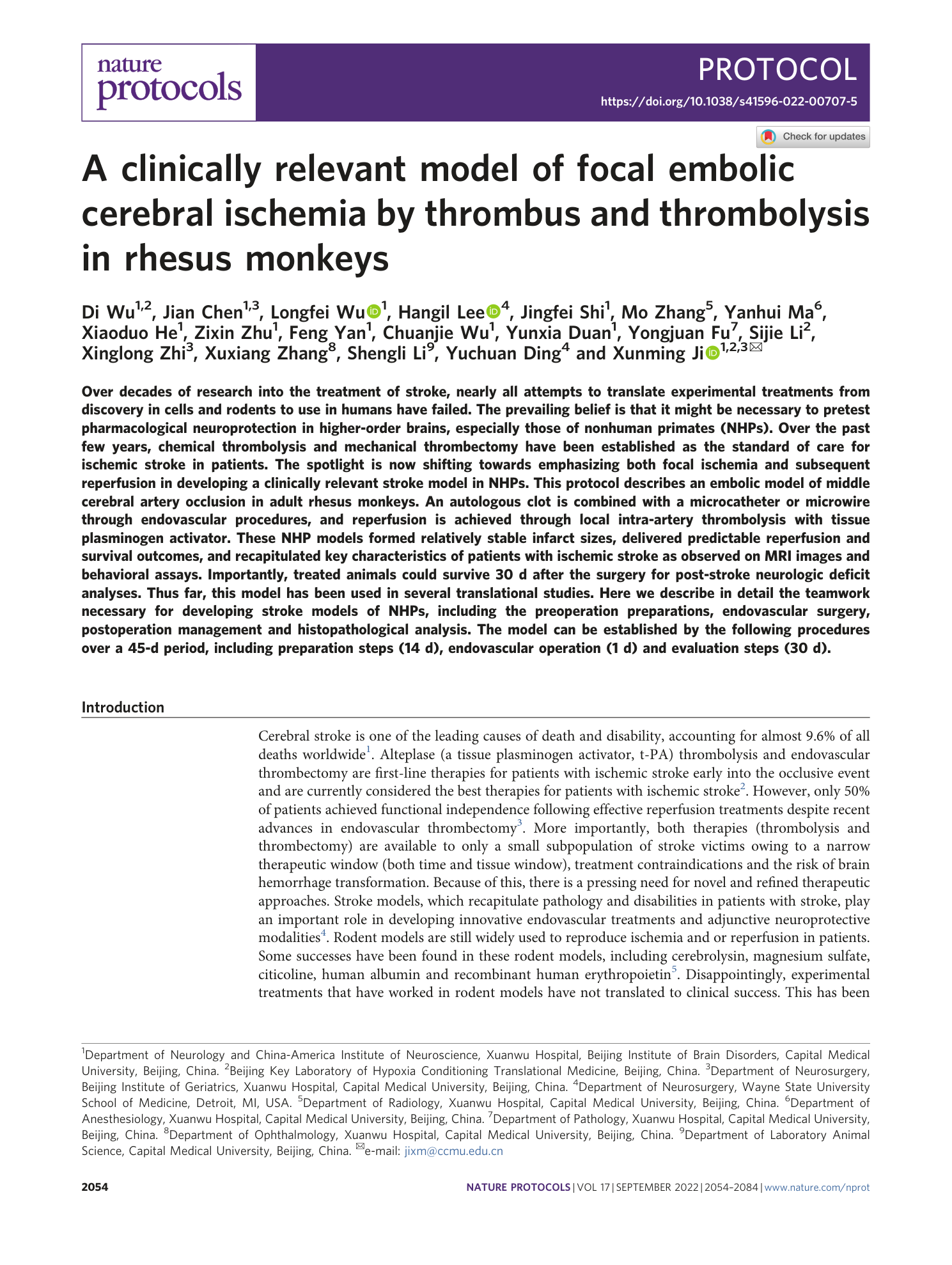
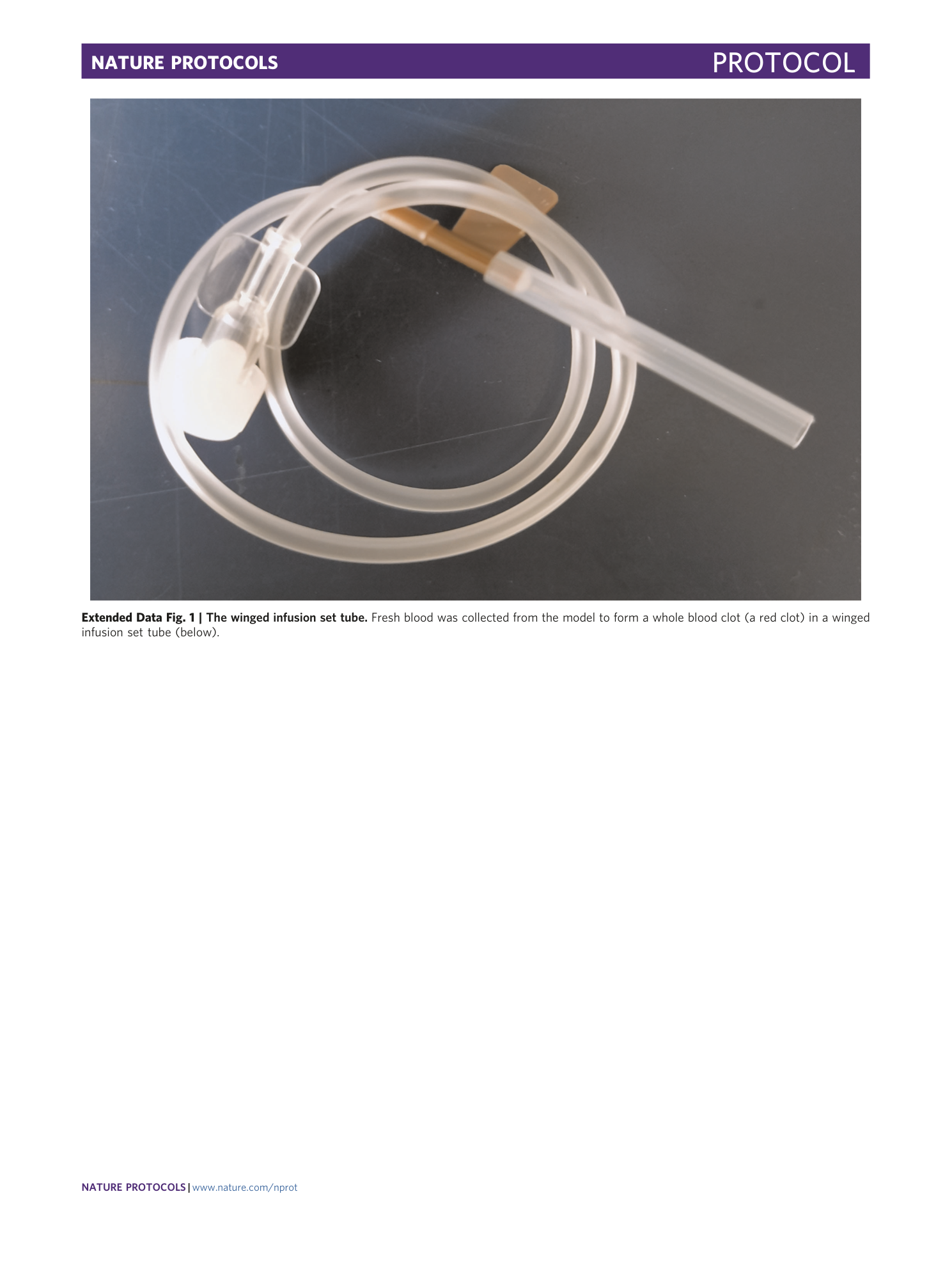
Extended Data Fig. 1 The winged infusion set tube.
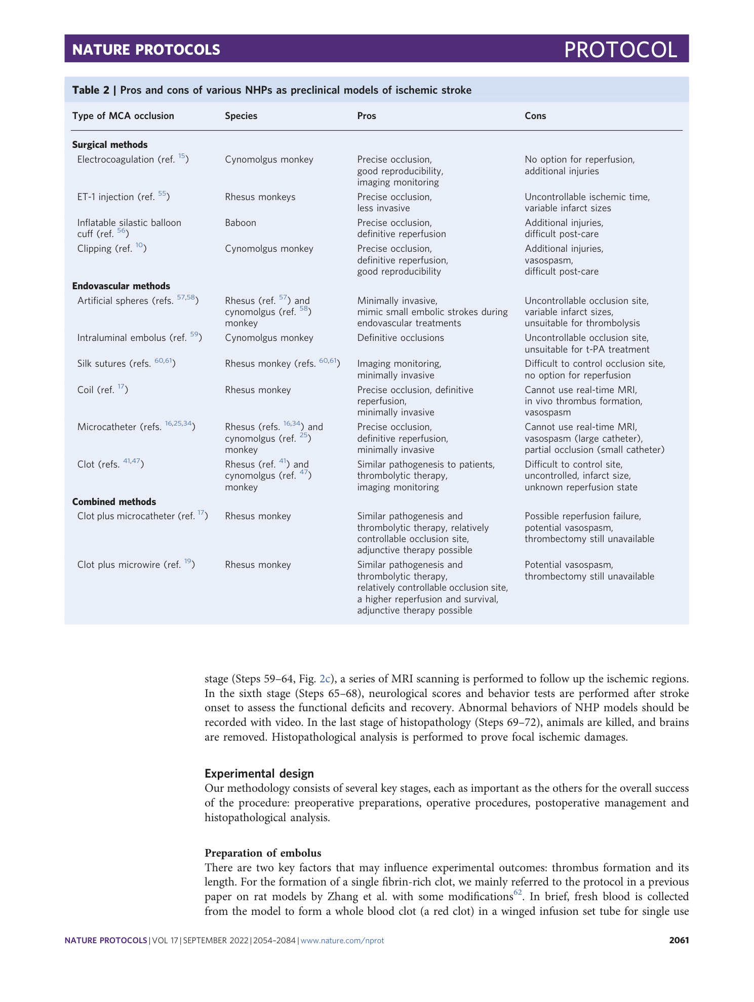
Fresh blood was collected from the model to form a whole blood clot (a red clot) in a winged infusion set tube (below).
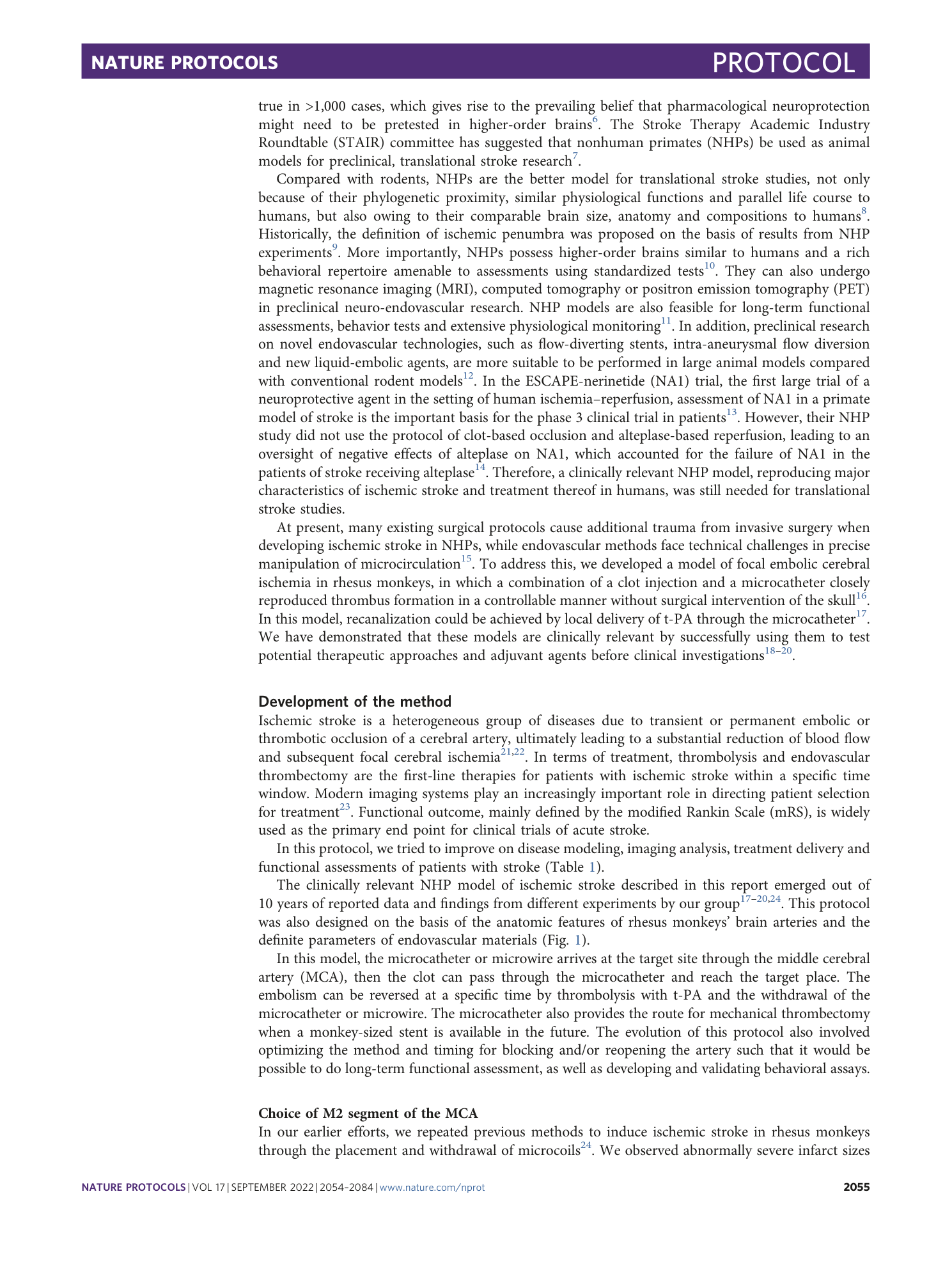
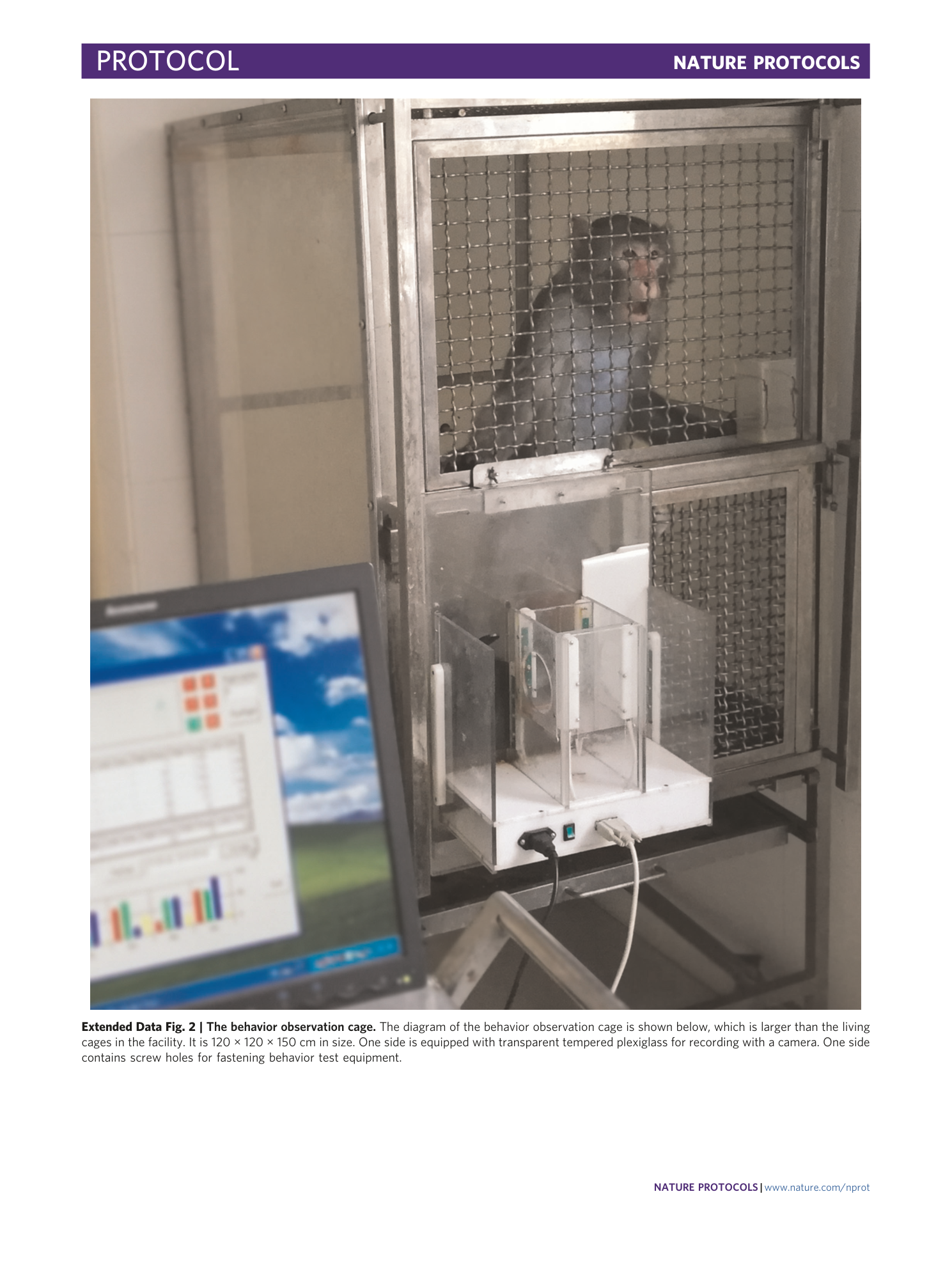
Extended Data Fig. 2 The behavior observation cage.
The diagram of the behavior observation cage is shown below, which is larger than the living cages in the facility. It is 120 × 120 × 150 cm in size. One side is equipped with transparent tempered plexiglass for recording with a camera. One side contains screw holes for fastening behavior test equipment.
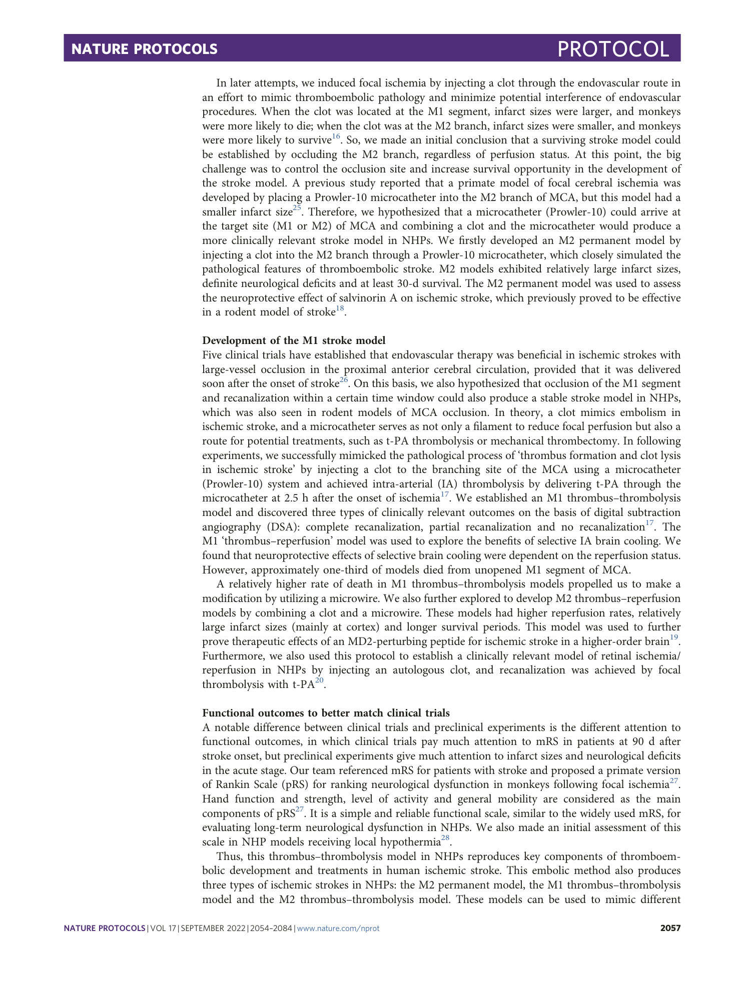
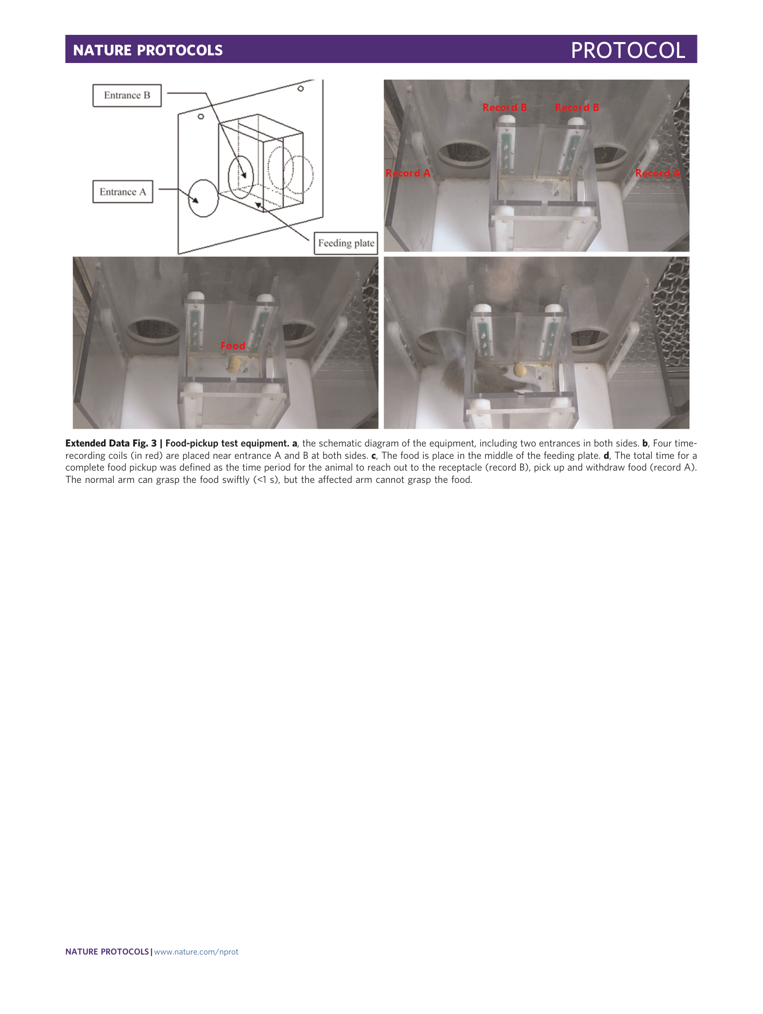
Extended Data Fig. 3 Food-pickup test equipment.
a , the schematic diagram of the equipment, including two entrances in both sides. b , Four time-recording coils (in red) are placed near entrance A and B at both sides. c , The food is place in the middle of the feeding plate. d , The total time for a complete food pickup was defined as the time period for the animal to reach out to the receptacle (record B), pick up and withdraw food (record A). The normal arm can grasp the food swiftly (<1 s), but the affected arm cannot grasp the food.
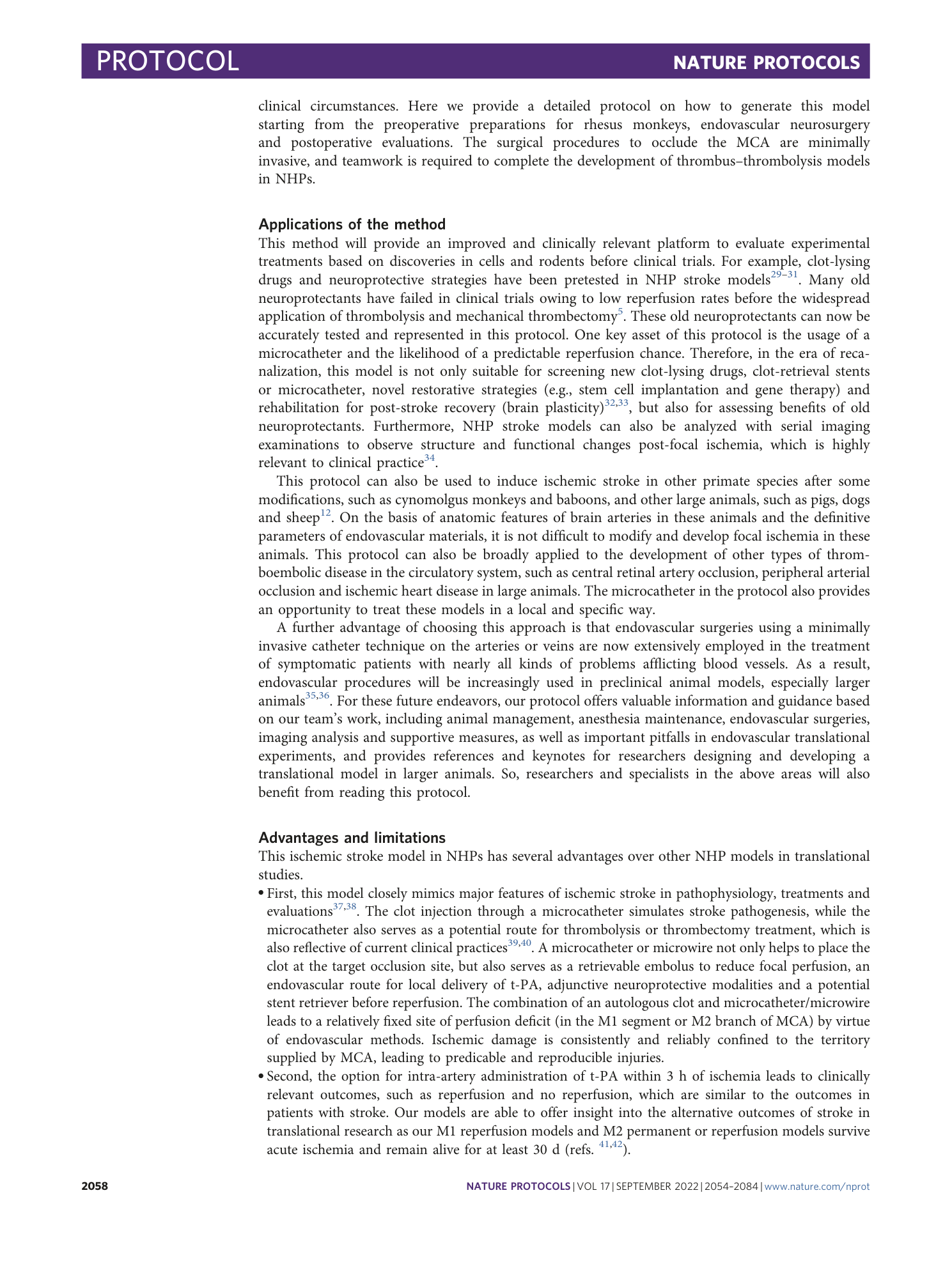
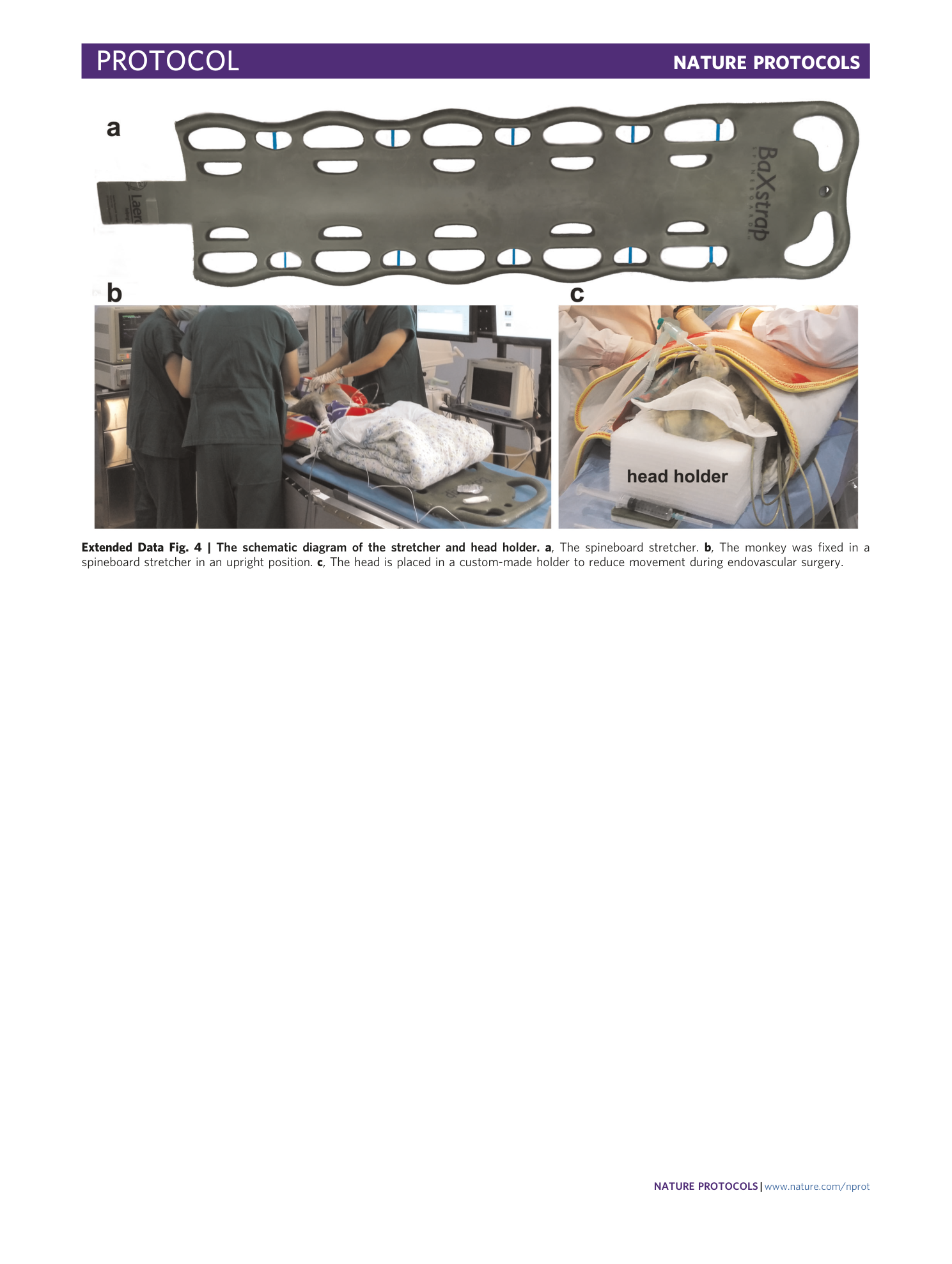
Extended Data Fig. 4 The schematic diagram of the stretcher and head holder.
a , The spineboard stretcher. b , The monkey was fixed in a spineboard stretcher in an upright position. c , The head is placed in a custom-made holder to reduce movement during endovascular surgery.
Extended Data Fig. 5 The impaired neurological functions over a 30-d observation period in a model of M1 occlusion and reperfusion.
Occlusion and reperfusion in the right MCA were achieved in this model. On the first day after stroke onset, the model was drowsy, showing no appetite for the fruit (1 in red) and no defense reaction. Grasp behavior was absent on the left side (2 in red), which was the damaged side. It did not walk or exhibit extremity movements (such as jump), and only crawled against the guardrail. On day 7, the monkey grasped the fruit (1 in red) with the nonaffected hand (right side), but the grasp was absent in the left hand (2 in red). It could crawl against the guardrail, showing minimal movement and profound weakness, without extremity movements. Facial weakness was profound with constant drooling (3 in red). On day 14, the monkey grasped the fruit (1 in red) with the nonaffected hand (right side) with some help from the affected hand (2 in red). The model exhibited a noticeable preference to turn to right side (circle in the clockwise direction, 4 in red). It could walk and sit on the rail (5 in red) and do some extremity movements (standing up and grasp the top rail, 6 in red). On day 30, the monkey grasped the fruit (1 in red) with the nonaffected hand (right side) with some help from the affected hand (2 in red), but the left arm and hand were noticeably impaired. The model could turn to left side (circle in the counterclockwise direction, 4 in red). It could do some extremity movements (standing up and grasp the top rail, 6 in red).
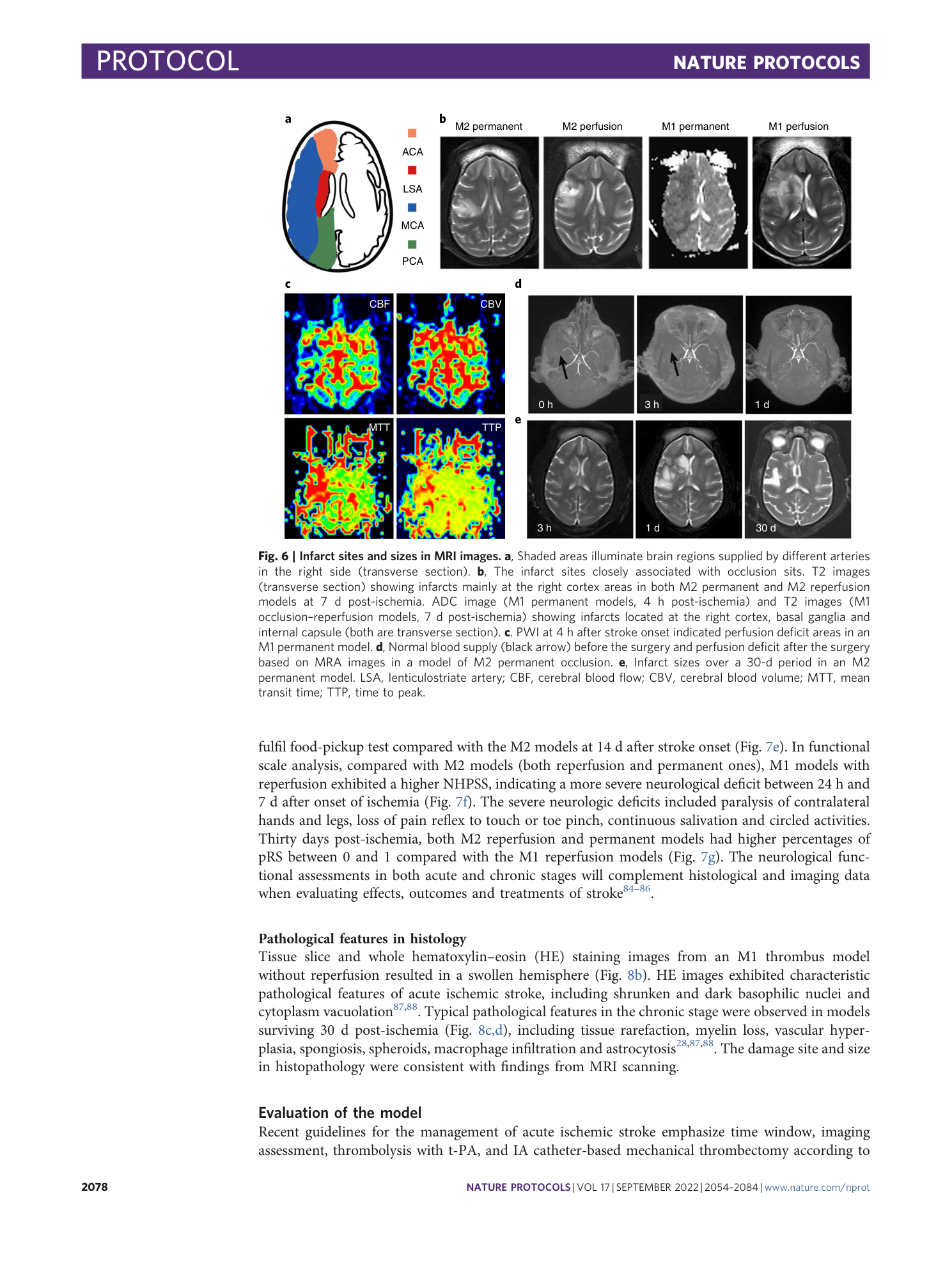
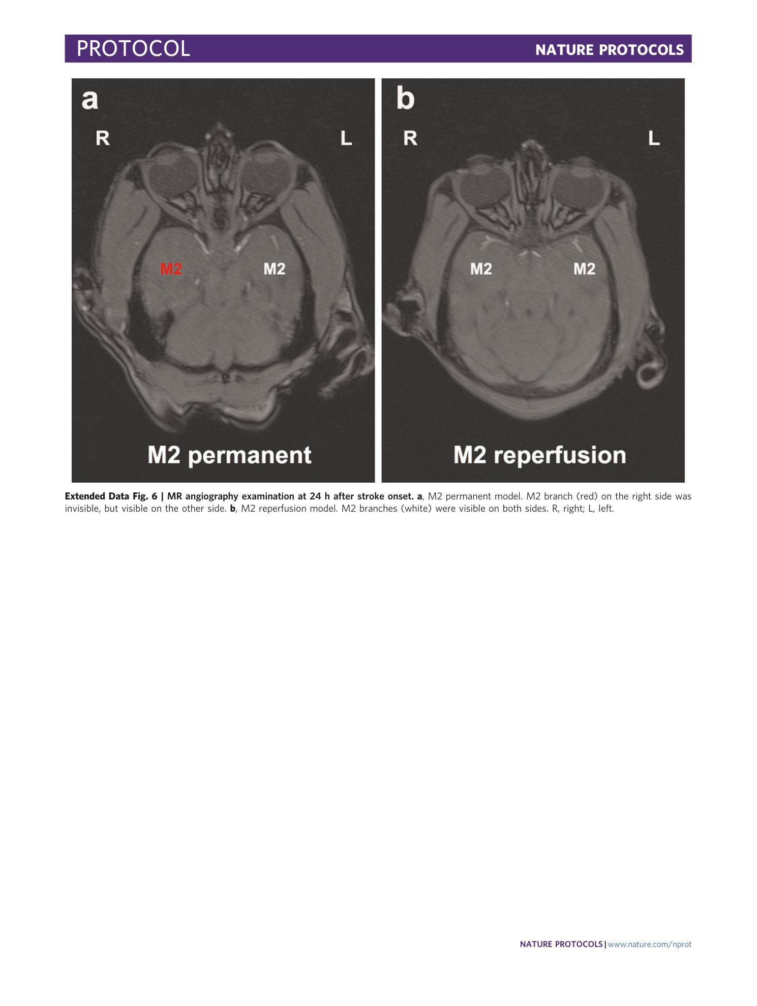
Extended Data Fig. 6 MR angiography examination at 24 h after stroke onset.
a , M2 permanent model. M2 branch (red) on the right side was invisible, but visible on the other side. b , M2 reperfusion model. M2 branches (white) were visible on both sides. R, right; L, left.
