Flow CyTOF Using Single Cells From Human Islets
Klaus H. Kaestner Lab, Suzanne Shapira
Abstract
Cytometry by time-of-flight (CyTOF) is an application of mass cytometry using antibodies conjugated to rare heavy metal isotopes combined with mass spectrometry. This technique significantly reduces background signals and spectral overlap often seen with traditional fluorescence methods. This protocol describes a technique to analyze multiple cellular features of human islets in parallel using a defined and optimized antibody cocktail.
Before start
Ι. Steps in pre-processing
-
Transfer handpicked islets (approximately 5,000 IEQs) into
15mLconical tube. Add10mLof 1xPBS w/o Ca²+, Mg²+ (Rockland, MB-008). Centrifuge for 2 min at RT, 180 xg. Aspirate the supernatant. -
Add
1mLof warm (37°C) 0.05% Trypsin (Invitrogen, 25300054) to the islets. Pipette up and down with p1000. -
Incubate at
37°Cfor 9 min, or until cells are in single cells. Pipette up and down at t=7 min, 4 min, 2 min, 0 min. Check under microscope towards the end of dissociation. -
Stop the trypsin reaction by adding
1mLof 100% FBS (Hyclone, SH3091003) to the disassembled islets and pass cells through BD FACs tube with strainer top (Corning 352235). Use1mLof 100% FBS to rinse the tube and pass through the strainer. -
Transfer cells to
15mLconical. Centrifuge 4 min, 400 xg. -
Dump out the supernatant and wash cells with PBS with 10% FBS. Centrifuge for 4 min, 400 xg.
-
Wash the cells with PBS and centrifuge for 4 min, 400 xg. Dump out the supernatant.
-
Cell counting. Resuspend cells to 1x10⁶/ml in PBS.
-
Cisplatin staining for live/dead differentiation. Dilute cisplatin stock (Fluidigm 201064) at 1:4000 in PBS. Incubate cells with cisplatin at RT 5 min.
Note: Time has to be exact since with time, cisplatin will enter live cell membranes.
-
Wash the cells with PBS+10% FBS.
-
Wash the cells again with PBS.
-
Fixation with 4% PFA at RT for 30 min. Make it fresh every time from 32% PFA (EMS 50-980-495).
Note: CyTOF has high demanding for fixation.
-
After fixing, spin at 800g x 4min.
-
Freeze at ~5M/500ul in Mr. Frosty.
Steps
II. CyTOF barcoding and labelling
Thaw the cells in quickly, and immediately add the Perm buffer 37°C quickly, and immediately add the Perm buffer
Remove the barcodes (Fluidigm 201060) from -20°C freezer and allow them to warm up to RT for at least 10 min.
Wash cells 2x with Foxp3 perm buffer (eBioscience, 00-5523-00), 700 xg (2900 rpm), 5min.
Note: centrifuge at higher speed after fixation.
Resuspend each sample to be barcoded in 800µL of perm buffer (aiming at 1 ~ 3x10⁶ cells)
Resuspend barcodes completely in 100µL of perm buffer and transfer them to the appropriate samples. Mix the samples immediately and completely (pipette immediately).
Incubate for 30 min at RT.
Centrifuge cells, wash twice with 2mL of perm buffer. 700 xg. 5 min.
Intracellular stain with master mix in perm +1% FBS at 4°C overnight. Master mix is 100ul/million for each tube.
Master Mix: 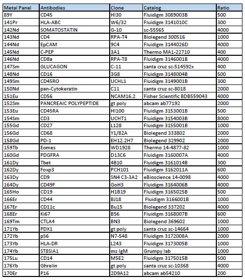
Wash with 2x perm buffer at 700 xg, 5min.
Wash 2x with PBS (700 xg, 5min)
DNA intercalator Iridium (FLuidigm 201192B, aliquoted in pcr tubes) at 1:4000 at RT for 1h in buffer to stain DNA is 2% PFA in PBS with Iridium.
Right before running samples on CyTOF2, wash (2x) with MilliQ H2O (700 xg, 5min)
Resuspend in MilliQ H2O. Adjust cell concentration to about 5x10⁵/ml (expect lose 40% of the starting cells at this step)
- alternatively count and resuspend to desired concentration.
Analyze on CyTOF at about 300-500 evt/sec
