Algorithmic assessment of cellular senescence in experimental and clinical specimens
J. Kohli, B. Wang, S. M. Brandenburg, N. Basisty, K. Evangelou, M. Varela-Eirin, J. Campisi, B. Schilling, V. Gorgoulis, M. Demaria
cellular senescence
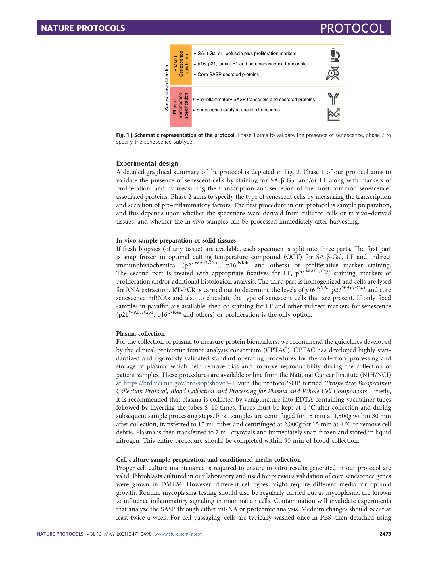
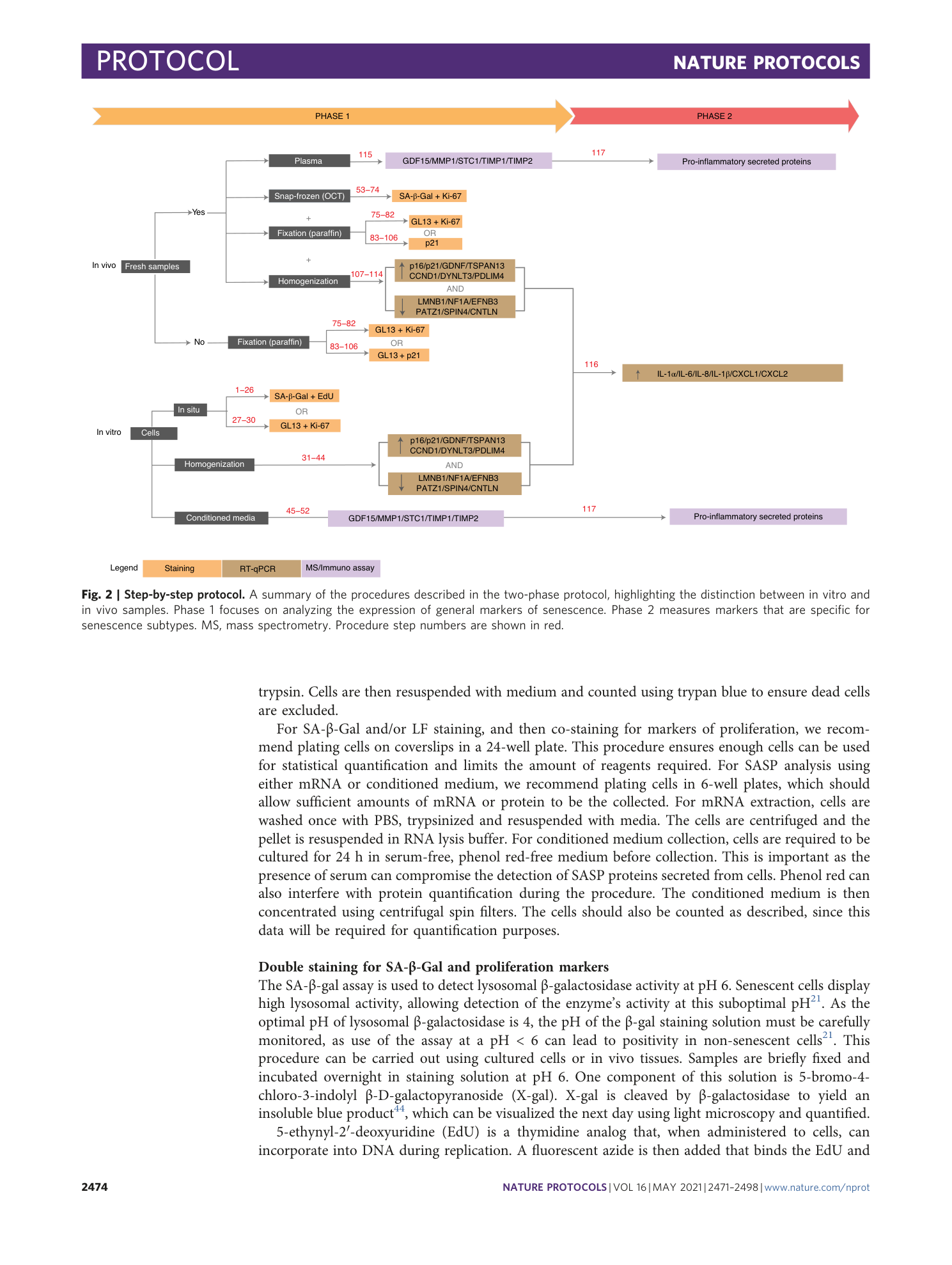
algorithmic assessment
senotherapies
senescence markers
phenotype characterization

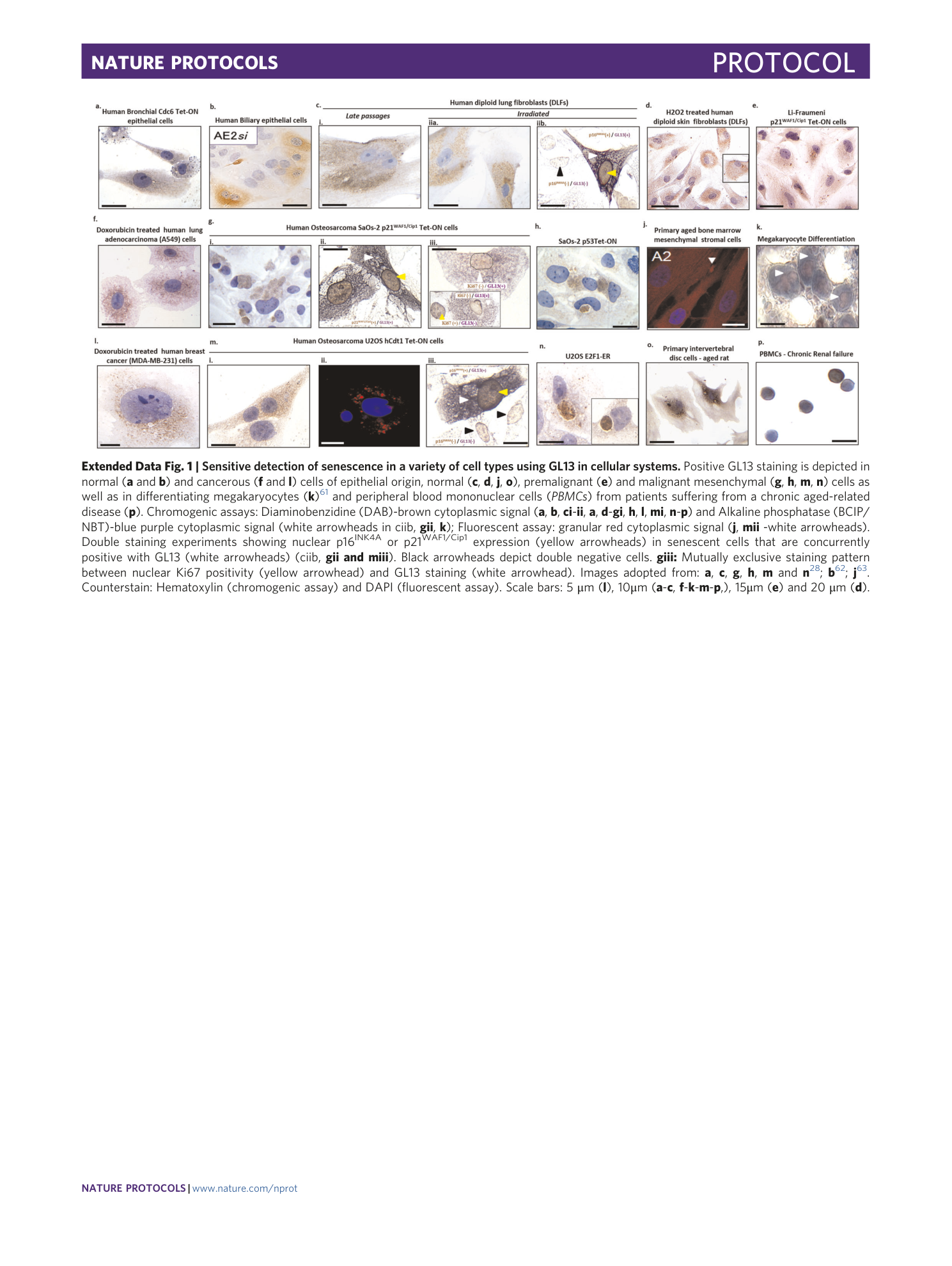
Extended
Extended Data Fig. 1 Sensitive detection of senescence in a variety of cell types using GL13 in cellular systems.
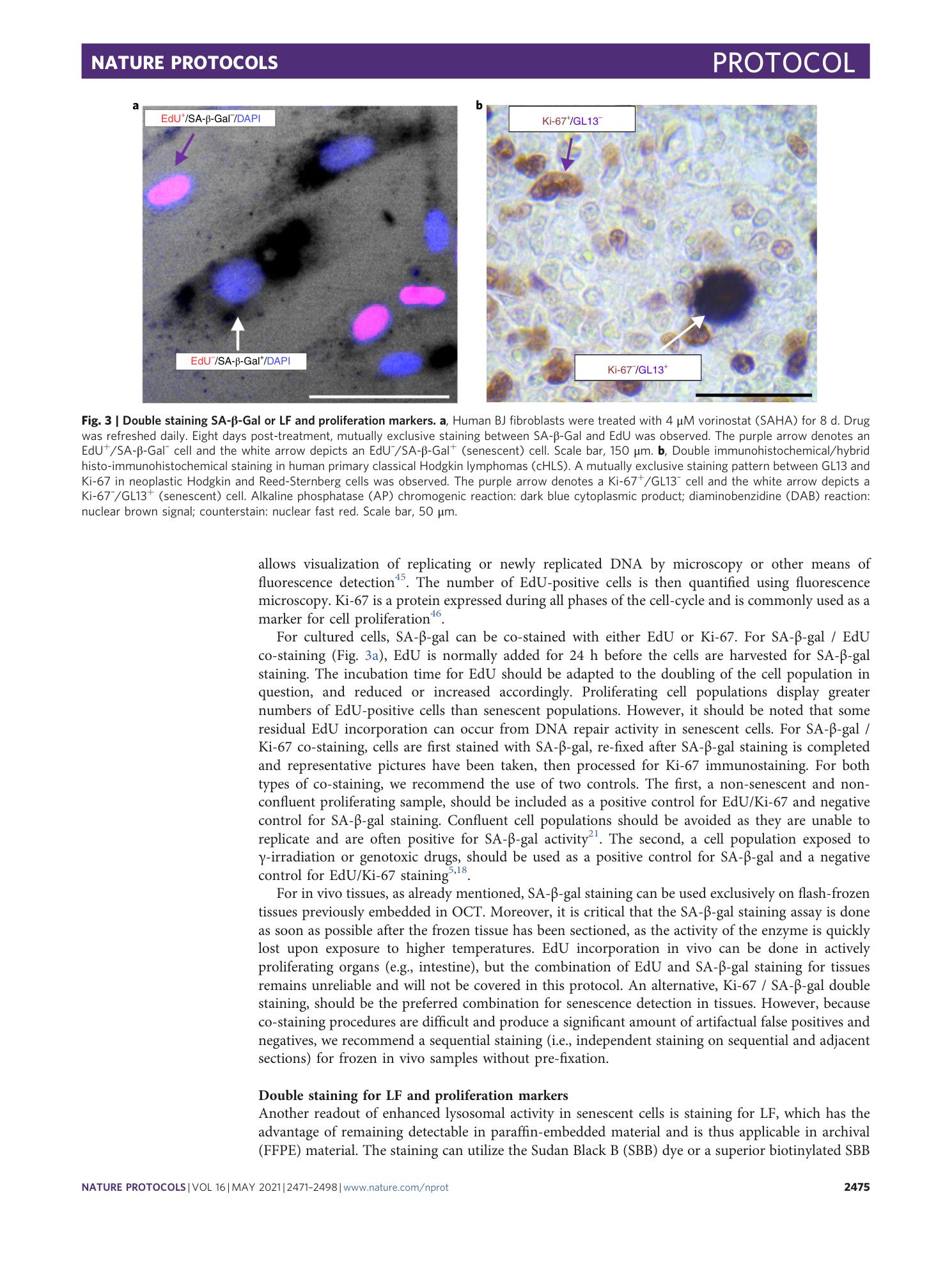
Positive GL13 staining is depicted in normal ( a and b ) and cancerous ( f and l ) cells of epithelial origin, normal ( c , d , j , o ), premalignant ( e ) and malignant mesenchymal ( g , h , m , n ) cells as well as in differentiating megakaryocytes ( k ) 61 and peripheral blood mononuclear cells ( PBMCs ) from patients suffering from a chronic aged-related disease ( p ). Chromogenic assays: Diaminobenzidine (DAB)-brown cytoplasmic signal ( a , b , ci - ii , a , d - gi , h , l , mi , n - p ) and Alkaline phosphatase (BCIP/NBT)-blue purple cytoplasmic signal (white arrowheads in ciib, gii , k ); Fluorescent assay: granular red cytoplasmic signal ( j , mii -white arrowheads). Double staining experiments showing nuclear p16 INK4A or p21 WAF1/Cip1 expression (yellow arrowheads) in senescent cells that are concurrently positive with GL13 (white arrowheads) (ciib, gii and miii ). Black arrowheads depict double negative cells. giii: Mutually exclusive staining pattern between nuclear Ki67 positivity (yellow arrowhead) and GL13 staining (white arrowhead). Images adopted from: a , c , g , h , m and n 28 ; b 62 ; j 63 . Counterstain: Hematoxylin (chromogenic assay) and DAPI (fluorescent assay). Scale bars: 5 μm ( l ), 10μm ( a - c , f - k - m - p ,), 15μm ( e ) and 20 μm ( d ).
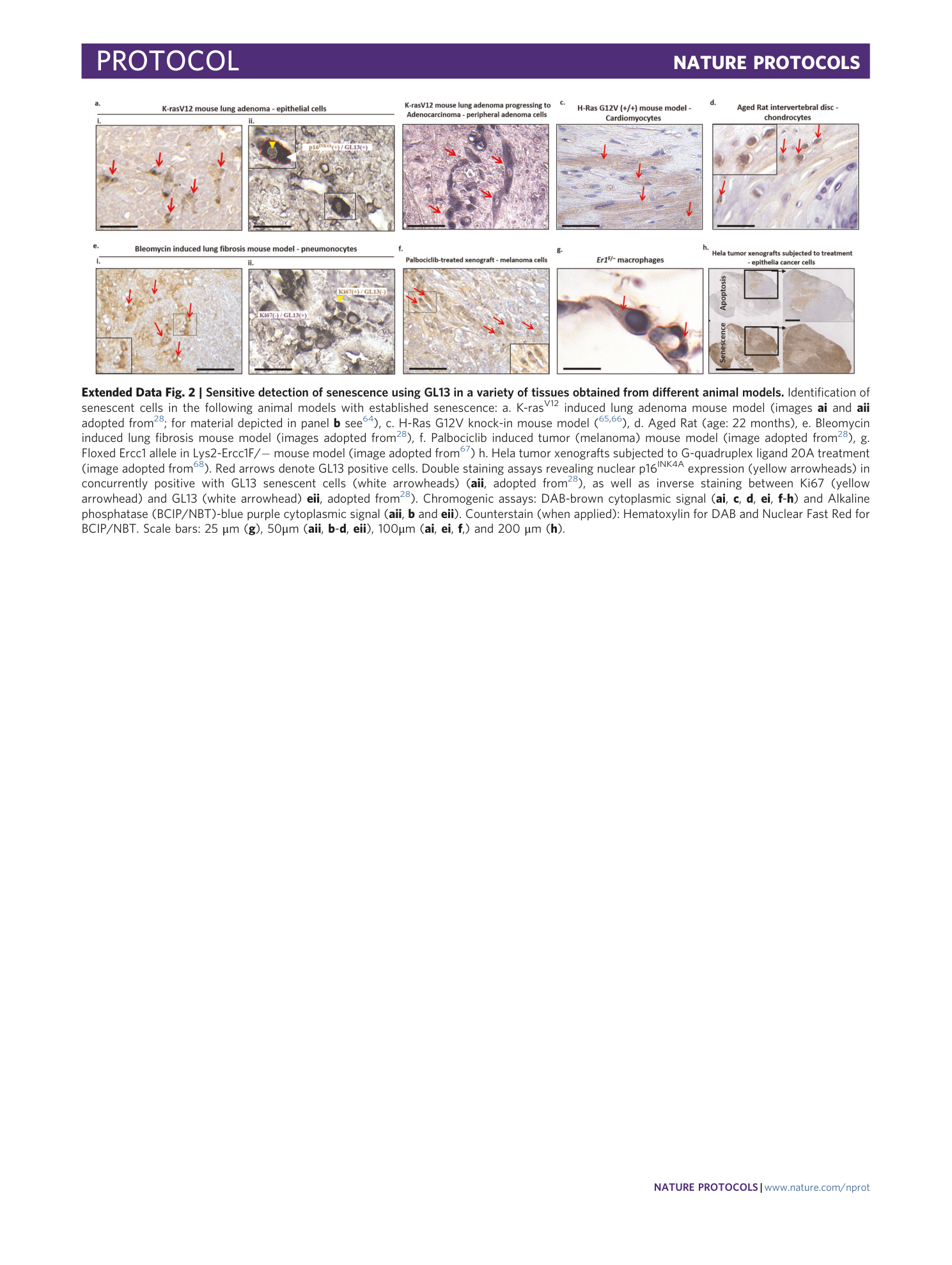
Extended Data Fig. 2 Sensitive detection of senescence using GL13 in a variety of tissues obtained from different animal models.
Identification of senescent cells in the following animal models with established senescence: a. K-ras V12 induced lung adenoma mouse model (images ai and aii adopted from 28 ; for material depicted in panel b see 64 ), c. H-Ras G12V knock-in mouse model ( 65 , 66 ), d. Aged Rat (age: 22 months), e. Bleomycin induced lung fibrosis mouse model (images adopted from 28 ), f. Palbociclib induced tumor (melanoma) mouse model (image adopted from 28 ), g. Floxed Ercc1 allele in Lys2-Ercc1F/− mouse model (image adopted from 67 ) h. Hela tumor xenografts subjected to G-quadruplex ligand 20A treatment (image adopted from 68 ). Red arrows denote GL13 positive cells. Double staining assays revealing nuclear p16 INK4A expression (yellow arrowheads) in concurrently positive with GL13 senescent cells (white arrowheads) ( aii , adopted from 28 ), as well as inverse staining between Ki67 (yellow arrowhead) and GL13 (white arrowhead) eii , adopted from 28 ). Chromogenic assays: DAB-brown cytoplasmic signal ( ai , c , d , ei , f - h ) and Alkaline phosphatase (BCIP/NBT)-blue purple cytoplasmic signal ( aii , b and eii ). Counterstain (when applied): Hematoxylin for DAB and Nuclear Fast Red for BCIP/NBT. Scale bars: 25 μm ( g ), 50μm ( aii , b - d , eii ), 100μm ( ai , ei , f ,) and 200 μm ( h ).
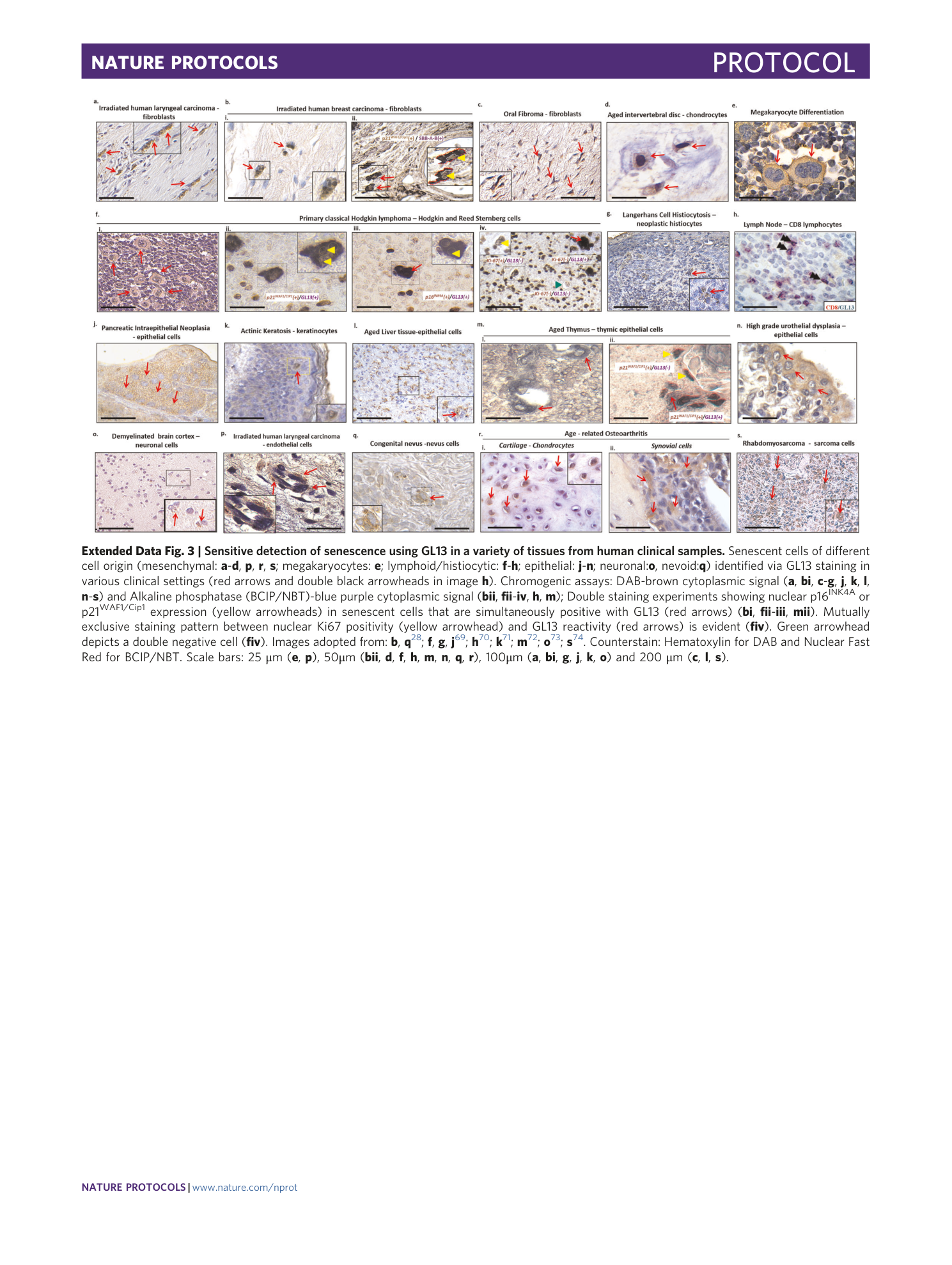
Extended Data Fig. 3 Sensitive detection of senescence using GL13 in a variety of tissues from human clinical samples.
Senescent cells of different cell origin (mesenchymal: a - d , p , r , s ; megakaryocytes: e ; lymphoid/histiocytic: f - h ; epithelial: j - n ; neuronal: o , nevoid: q ) identified via GL13 staining in various clinical settings (red arrows and double black arrowheads in image h ). Chromogenic assays: DAB-brown cytoplasmic signal ( a , bi , c - g , j , k , l , n - s ) and Alkaline phosphatase (BCIP/NBT)-blue purple cytoplasmic signal ( bii , fii - iv , h , m ); Double staining experiments showing nuclear p16 INK4A or p21 WAF1/Cip1 expression (yellow arrowheads) in senescent cells that are simultaneously positive with GL13 (red arrows) ( bi , fii - iii , mii ). Mutually exclusive staining pattern between nuclear Ki67 positivity (yellow arrowhead) and GL13 reactivity (red arrows) is evident ( fiv ). Green arrowhead depicts a double negative cell ( fiv ). Images adopted from: b , q 28 ; f , g , j 69 ; h 70 ; k 71 ; m 72 ; o 73 ; s 74 . Counterstain: Hematoxylin for DAB and Nuclear Fast Red for BCIP/NBT. Scale bars: 25 μm ( e , p ), 50μm ( bii , d , f , h , m , n , q , r ), 100μm ( a , bi , g , j , k , o ) and 200 μm ( c , l , s ).
