Synthesis and functionalization of scalable and versatile 2D protein films via amyloid-like aggregation
Yongchun Liu, Shuting Miao, Hao Ren, Lihua Tian, Jian Zhao, Peng Yang


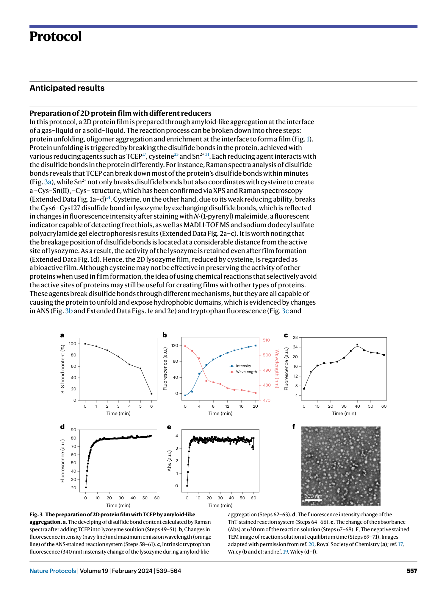


Extended
Extended Data Fig. 1 2D lysozyme film prepared with Sn 2+ .
a - c , Survey (a) and high-resolution XPS spectra of Sn 3d (b) and S 2p (c) of the film. d , Raman spectra of native lysozyme and film, with the inset showing the characteristic peaks for the clusters of -S-(Sn)x-S- at 309 cm-1 and reduction of the disulfide bond at 505 cm-1, respectively. e , Evolution of ANS fluorescence to monitor the exposition of hydrophobic domain during amyloid-like aggregation. f , The fluorescence intensity of the ThT-stained reaction solution at different aggregation time. Figure adapted with permission from ref. 31 , ACS.
Extended Data Fig. 2 2D lysozyme film prepared with cysteine.
a , The NPM assay for native lysozyme and partially unfolded lysozyme. The inset shows the reaction between NPM and free thiol to enhance the fluorescence emission. b , Typical MALDI-TOF-MS spectra of native lysozyme and cysteine-reduced lysozyme. c , The SDS-PAGE of native lysozyme (lane 1) and partially unfolded lysozyme (lane 2). d ,The activity assay of native lysozyme, cysteine and partially unfolded lysozyme. e , ANS assay for native lysozyme, cysteine, and partially unfolded lysozyme. f , Intrinsic tryptophan fluorescence assay for native lysozyme and partially unfolded lysozyme. Figure adapted with permission from ref. 23 , Wiley.
Extended Data Fig. 3 Characterization of the 2D lysozyme film prepared with Sn 2+ .
a , Digital photograph showing a large-area protein film at the air/water interface. b , Scanning transmission electron microscopy (STEM) image of the protein film. c , Corresponding energy-dispersive X-ray spectroscopy (EDS) elemental mapping of Sn in the protein film. d , Water contact angle measurements of the protein film on different substrates. e , Transmittance of the protein film recorded by UV/vis spectrometer. f , Fluorescence spectra of the protein film stained with ThT. g , CLSM image of the ThT-dyed protein film. h ,Digital photograph of the protein film dyed with Congo red. i , Film thickness as a function of lysozyme concentration. Figure adapted with permission from ref. 31 , ACS.
Extended Data Fig. 4 Characterization of the 2D lysozyme film prepared with cysteine.
a , Digital photograph of a large-area protein film at the air/water interface. b , c , AFM (b) and TEM (c) images of the protein film surface. d , UV/vis absorption spectrum of the protein film in the range of 200-800 nm, along with a digital photograph showing the film-coated quartz with a leaf as a background. e , Water contact angle of the protein film coated on different substrates. f , CD spectra of the protein film stained with ThT. g , Digital photograph of the protein film dyed by Congo Red and an CLSM image of the ThT-dyed protein film. h , The thickness of the film as a function of lysozyme concentration. i , The effect of repetitive coating times on the thickness of the nanofilm. Figure adapted with permission from ref. 23 , Wiley.
Supplementary information
Supplementary Information
Supplementary Figs. 1–6 and Tables 1 and 2.
Reporting Summary
Supplementary Data 1
Statistical source data for Supplementary Fig. 5b–d.
