Immunoassay of SARS-CoV-2 in dogs and cats V.1
Dumar A. Jaramillo-Hernández, María C. Chacón, María A. Velásquez, Adolfo Vásquez-Trujillo, Ana P. Sánchez, Luis F. Salazar Garces, Gina L. García, Yohana M. Velasco-Santamaría, Luz N. Pedraza, Lida C. Lesmes-Rodríguez
Abstract
This protocol describes the qualitative determination of IgG antibodies against the nucleocapsid (N) protein of SARS-CoV-2 in serum from domestic dogs and cats, the indirect ELISA (Enzyme-Linked Immunosorbent Assay) kit ID Screen® SARS-CoV-2 Double Antigen Multi-Species (IDvet, Grabels, France).
The diagnostic kit detects antibodies against the nucleocapsid (N protein) of the SARS-CoV-2 virus.
This test can be used on samples from dogs, cats, mink, ferrets, cattle, sheep, goats, horses and any other susceptible species requiring serum, plasma or whole blood samples.
Steps
Before you start
Collect the serum, plasma or whole blood samples from dogs, cats, mink, ferrets, cattle, sheep, goats, horses and any other susceptible species.
Procedure
Allow the reagents to reach room temperature (21 °C ± 5 °C) before use.
Homogenize all reagents and serum samples by immersion or vortexing.
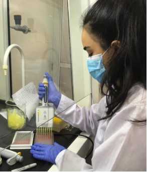
Add 25 µL of Negative control to wells A1 and A2 and 25 µL of Positive control to wells A3 and A4.
Add 25 µL of Diluent 13 to the remaining wells.
Cover the plate and incubate for 45 minutes ± 5 minutes at 37 °C ( ± 2°C).
Empty the wells and wash each well 5 times with at least 300 µL of wash solution, avoid drying the wells between washes.
Prepare the 1X conjugate by diluting the 10X conjugate 1:10 with diluent 13.
Add 100 µL of the 1X conjugate to each well.
Cover the plate and incubate for 30 minutes at 21°C (± 5°C).
Empty the wells.
Add at least 300 µL of wash solution so that this substrate reacts with the secondary antibody enzyme and provides a visible signal that can be quantified.
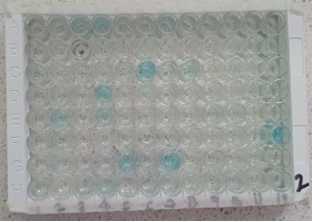
Add 100 µL of the developing solution into each well.
Cover the plate and incubate 20 min ± 2 min at 21°C (± 5°C) in the dark.
Finally, the optical density (OD) is read in a Cytation 3 multimodal microplate reader (BioTek Instruments, Inc. Winooski, VT, USA) using a wavelength of 450 nm.
Validation
The assay is validated if:
- The mean value of the Positive Control O.D. (DOcp) is greater than 0.350.
DOcp > 0.350
- The ratio of the mean of the optical density values of the positive and negative controls (DOcp and DOcn) is greater than 3.
DOcp/DOcn > 3
Interpretation
After the laboratory reading, the sample/positive control (S/P) ratio was calculated with the data obtained, which was expressed as a percentage, using the following formula:
Samples presenting a S/P%:
-
Less than or equal to 50% are considered negative.
-
Between 50% and 60% are considered doubtful.
-
Greater than or equal to 60% are considered positive.
Results Status
S/P % ≤ 50 % Negative
50 % < S/P % < 60% Doubtful
S/P % ≥ 60 % Positive


