MTT (Assay protocol
Abdulkareem Hameed Abd, Enas Jawad Kadhim, Matin Mahmood
Abstract
MTT (( 3-(4,5-Dimethylthiazol-2-yl)-2,5-Diphenyltetrazolium Bromide )) is used to assess cell viability as a function of redox potential. Actively respiring cells convert the water-soluble MTT to an insoluble purple formazan.
AMJ13: 2 (Ahmed.Murtuda ,Jabria 2013)cell line: 2 (Ahmed.Murtuda ,Jabria 2013)cell line:
The cell line of breast cancer has been obtained from Iraqi breast cancer which originated from the prime tumor of an old Iraqi woman (70 years) with a histological identification with carcinoma of infiltrating ductal (1).
SK-GT-4: esophageal carcinomacell line was established from a primary tumors in 1989 from a 89 year-old Caucasian male who presented with dysphagia secondary to a well-differentiated adenocarcinoma arising in the Barrett epithelium of the distal oesophagus
Steps
Maintenance of Cell lines
SK-GT-4 cell line, was maintained in MEM supplemented with 10% Fetal bovine, 100 units/mL penicillin, and 100 µg/mL streptomycin. Cells were passaged using Trypsin-EDTA reseeded at 50% confluence twice a week, and incubated at 37 °C
<NHF cell line, was maintained in MEM supplemented with 10% Fetal bovine, 100 units/mL penicillin, and 100 µg/mL streptomycin. Cells were passaged using Trypsin-EDTA reseeded at 50% confluence twice a week, and incubated at 37 °C >
MTT Assay
To determine the cytotoxic effect, the MTT cell viability assay was conducted on 96-well plates
Cell lines were seeded at 1 × 10⁴cells/well. After 24 hrs. or a confluent monolayer was achieved, cells were treated with tested compound
Cell viability was measured after 72h of treatment by removing the medium
adding 28 µL of 2 mg/mL solution of MTT
incubating the cells for 1.5 h at 37 °C.
After removing the MTT solution, the crystals remaining in the wells were solubilized by the addition of 130 µL of DMSO (Dimethyl Sulphoxide)
followed by 37 °C incubation for 15 min with shaking (orbital shaker)
The absorbency was determined on a microplate reader at 492 nm (test wavelength)
The assay was performed in triplicate
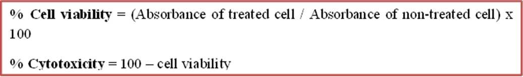
The inhibition rate of cell growth (the percentage of cytotoxicity) was calculated as the following equation