Isolation of mitochondria-derived mitovesicles and subpopulations of microvesicles and exosomes from brain tissues
Pasquale D’Acunzo, Yohan Kim, Jonathan M. Ungania, Rocío Pérez-González, Chris N. Goulbourne, Efrat Levy
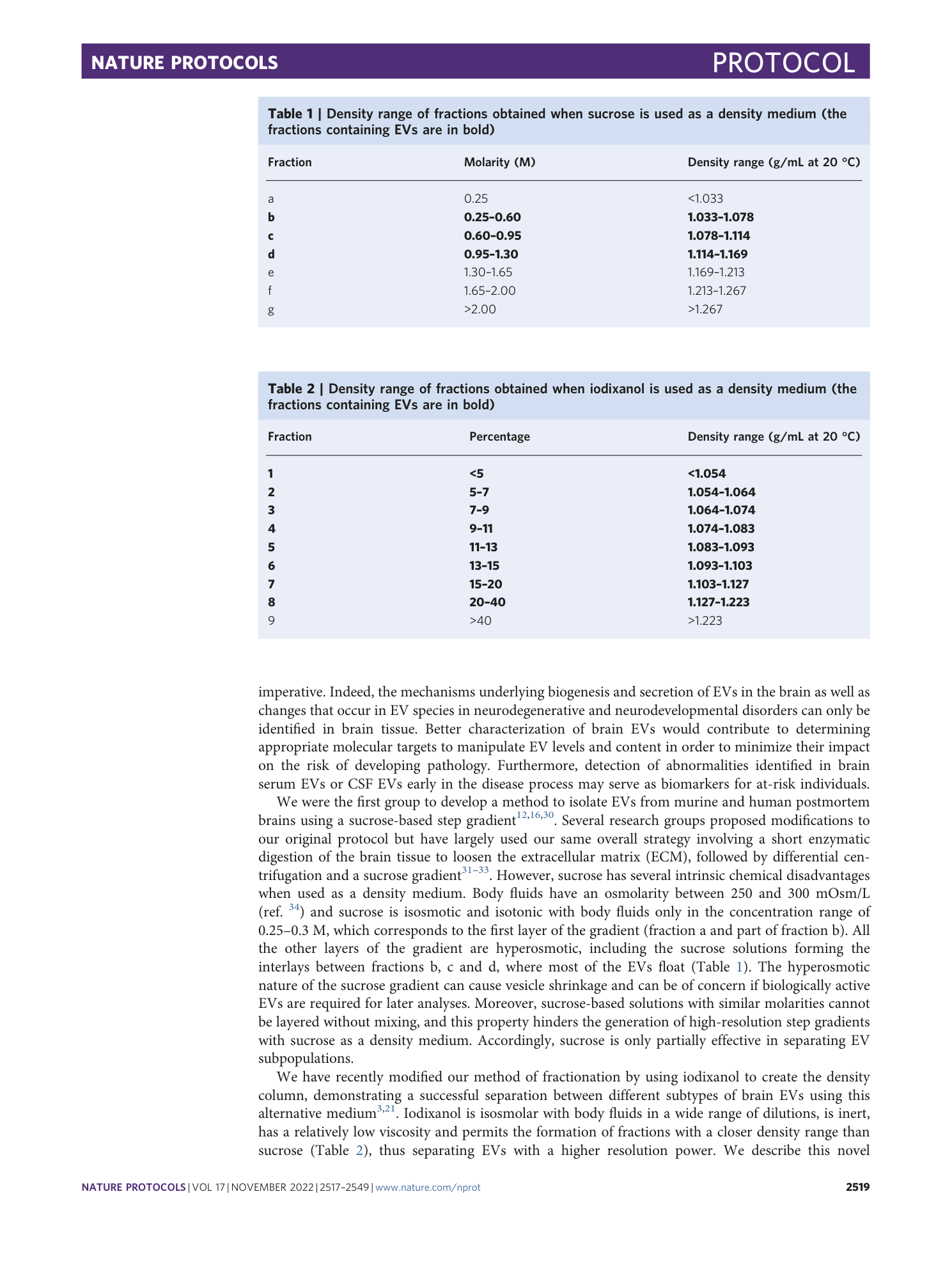
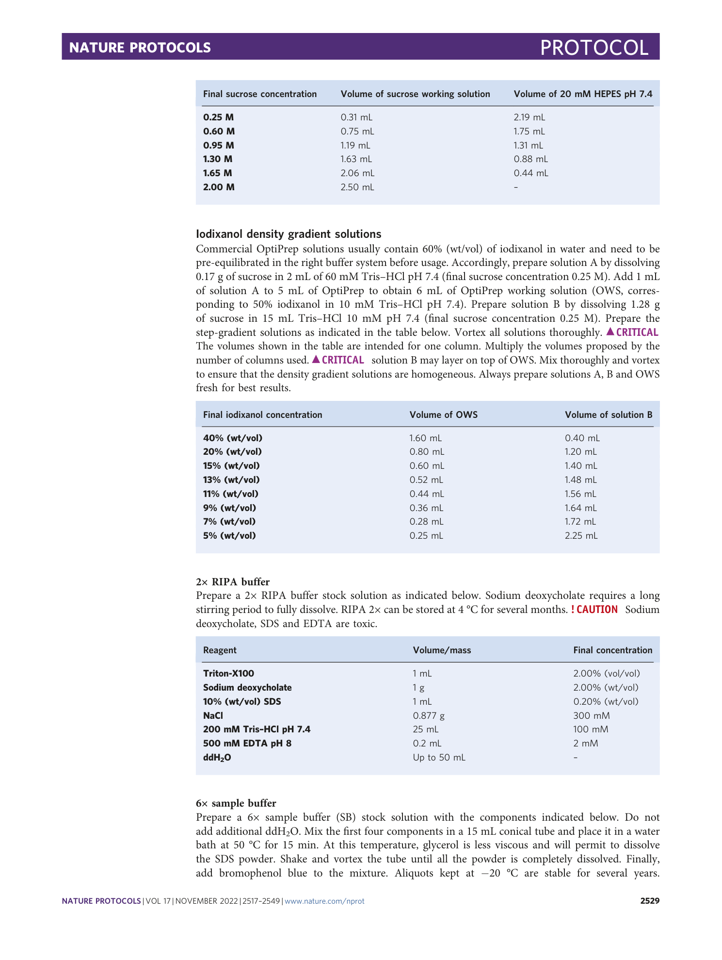

Extended
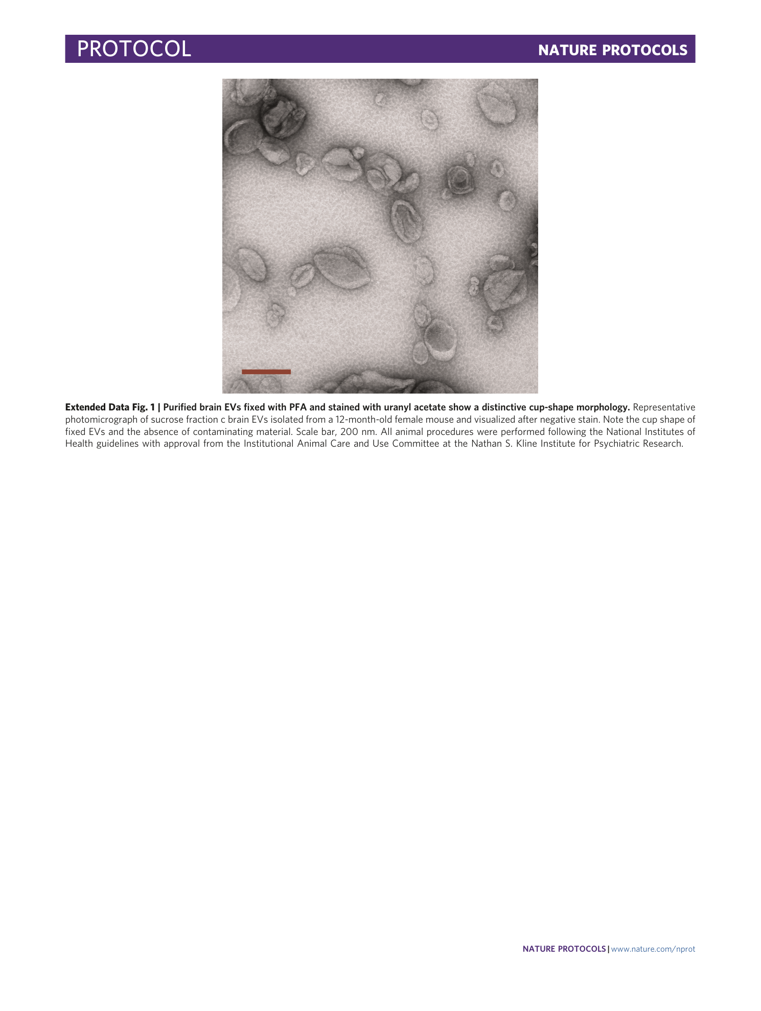
Extended Data Fig. 1 Purified brain EVs fixed with PFA and stained with uranyl acetate show a distinctive cup-shape morphology.
Representative photomicrograph of sucrose fraction c brain EVs isolated from a 12-month-old female mouse and visualized after negative stain. Note the cup shape of fixed EVs and the absence of contaminating material. Scale bar, 200 nm. All animal procedures were performed following the National Institutes of Health guidelines with approval from the Institutional Animal Care and Use Committee at the Nathan S. Kline Institute for Psychiatric Research.
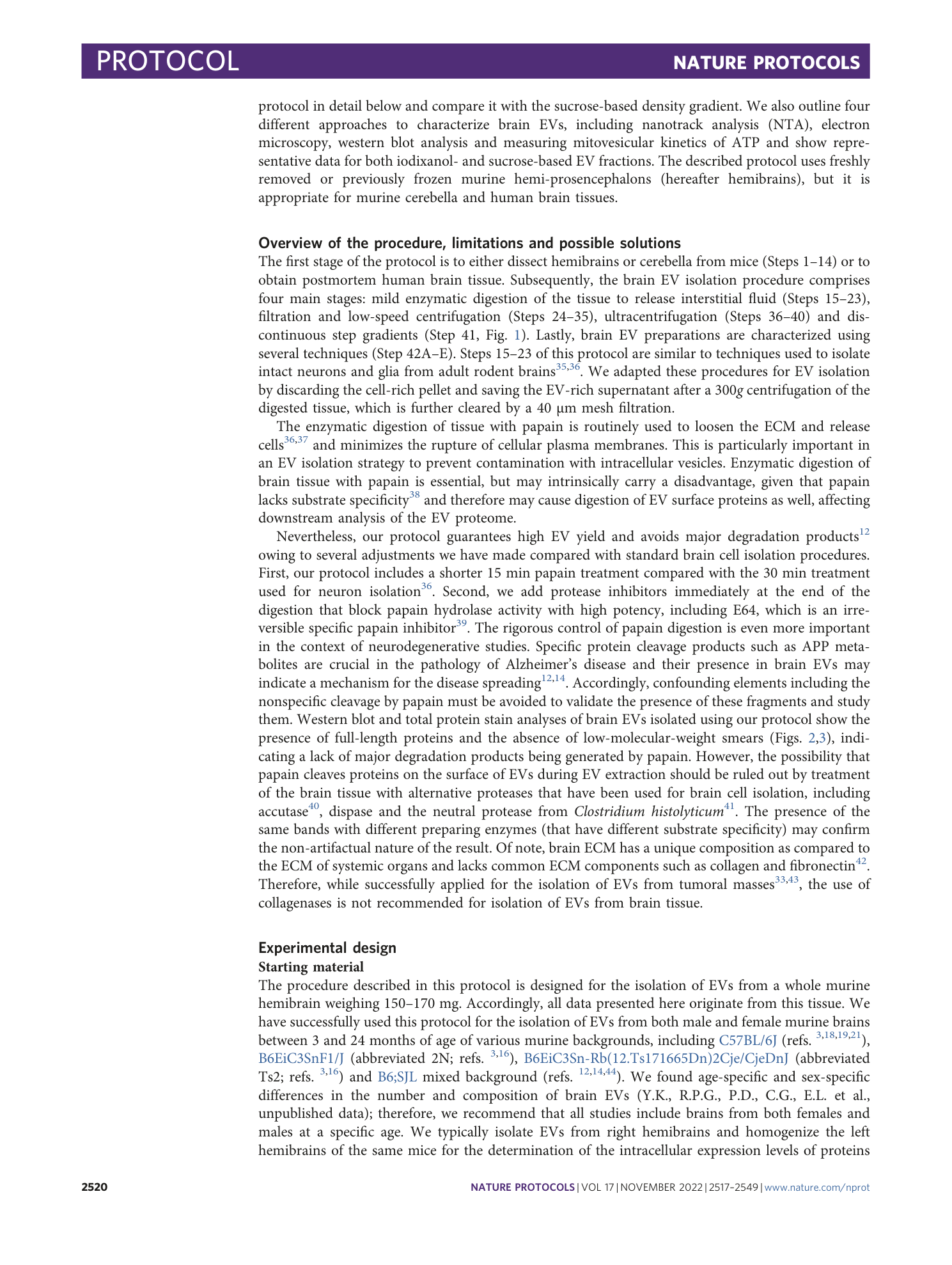
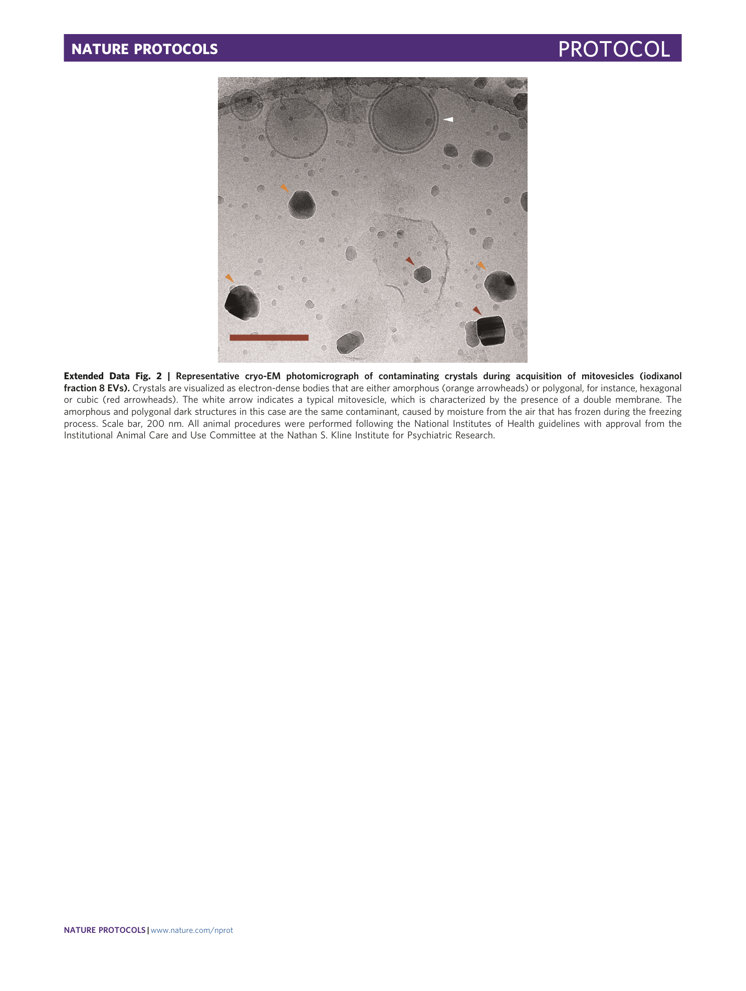
Extended Data Fig. 2 Representative cryo-EM photomicrograph of contaminating crystals during acquisition of mitovesicles (iodixanol fraction 8 EVs).
Crystals are visualized as electron-dense bodies that are either amorphous (orange arrowheads) or polygonal, for instance, hexagonal or cubic (red arrowheads). The white arrow indicates a typical mitovesicle, which is characterized by the presence of a double membrane. The amorphous and polygonal dark structures in this case are the same contaminant, caused by moisture from the air that has frozen during the freezing process. Scale bar, 200 nm. All animal procedures were performed following the National Institutes of Health guidelines with approval from the Institutional Animal Care and Use Committee at the Nathan S. Kline Institute for Psychiatric Research.
