CODEX multiplexed tissue imaging with DNA-conjugated antibodies
Christian M. Schürch, Sarah Black, Darci Phillips, John W. Hickey, Julia Kennedy-Darling, Vishal G. Venkataraaman, Nikolay Samusik, Yury Goltsev, Garry P. Nolan


Extended
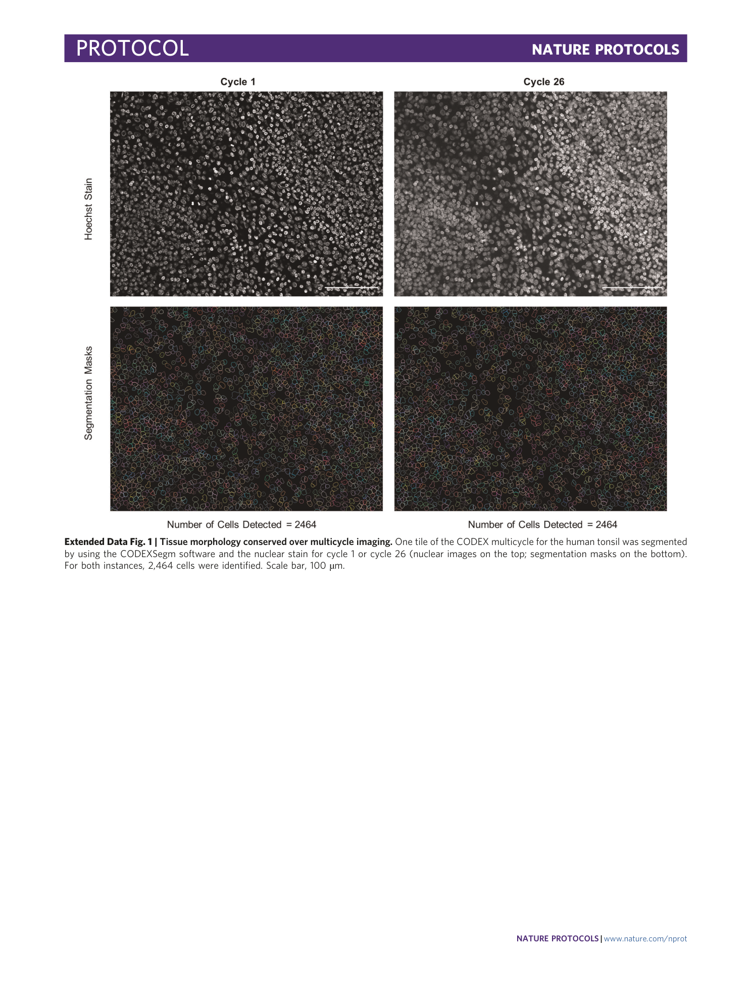
Extended Data Fig. 1 Tissue morphology conserved over multicycle imaging.
One tile of the CODEX multicycle for the human tonsil was segmented by using the CODEXSegm software and the nuclear stain for cycle 1 or cycle 26 (nuclear images on the top; segmentation masks on the bottom). For both instances, 2,464 cells were identified. Scale bar, 100 µm.
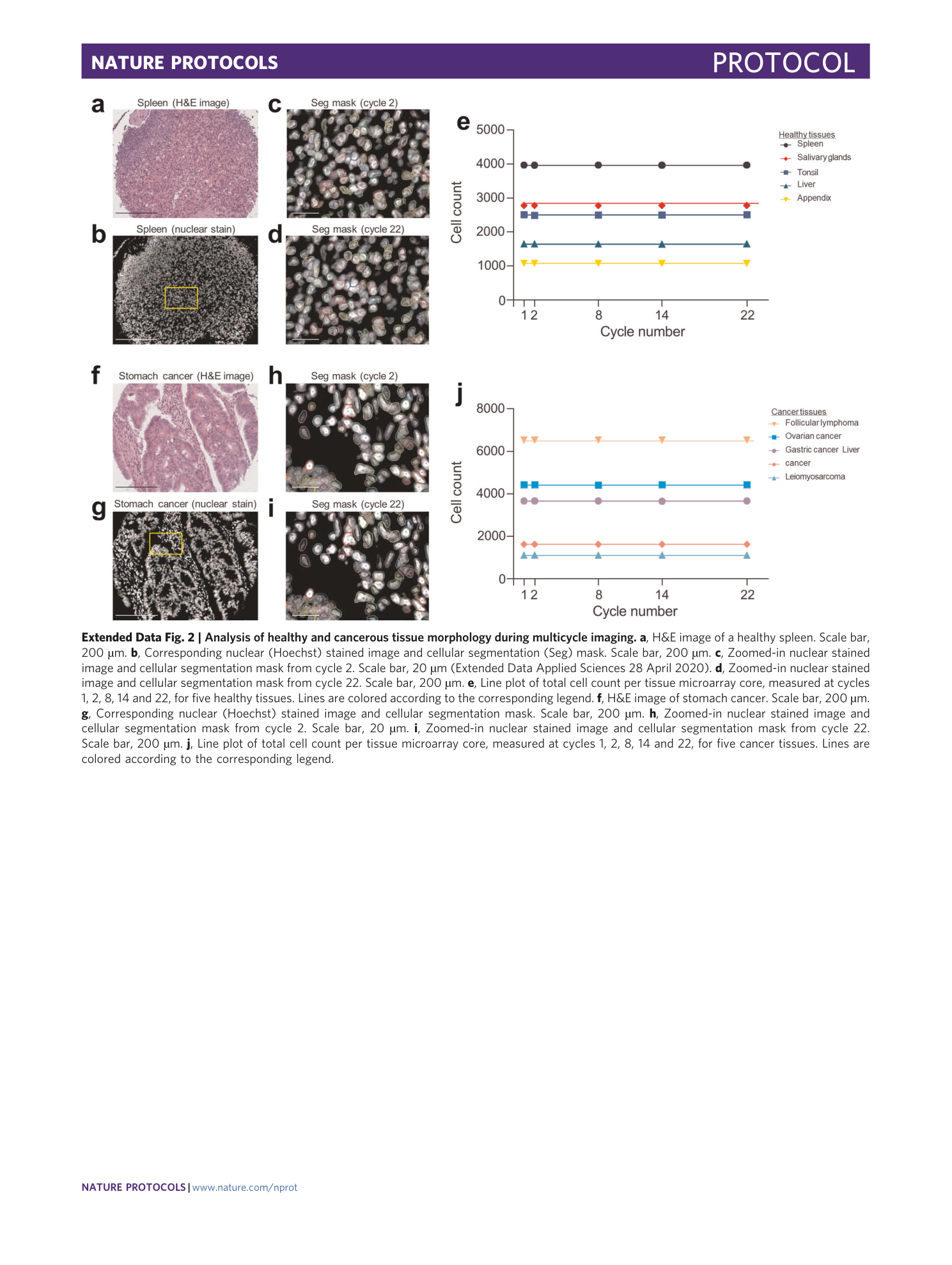
Extended Data Fig. 2 Analysis of healthy and cancerous tissue morphology during multicycle imaging.
a , H&E image of a healthy spleen. Scale bar, 200 µm. b , Corresponding nuclear (Hoechst) stained image and cellular segmentation (Seg) mask. Scale bar, 200 µm. c , Zoomed-in nuclear stained image and cellular segmentation mask from cycle 2. Scale bar, 20 µm (Extended Data Applied Sciences 28 April 2020). d , Zoomed-in nuclear stained image and cellular segmentation mask from cycle 22. Scale bar, 200 µm. e , Line plot of total cell count per tissue microarray core, measured at cycles 1, 2, 8, 14 and 22, for five healthy tissues. Lines are colored according to the corresponding legend. f , H&E image of stomach cancer. Scale bar, 200 µm. g , Corresponding nuclear (Hoechst) stained image and cellular segmentation mask. Scale bar, 200 µm. h , Zoomed-in nuclear stained image and cellular segmentation mask from cycle 2. Scale bar, 20 µm. i , Zoomed-in nuclear stained image and cellular segmentation mask from cycle 22. Scale bar, 200 µm. j , Line plot of total cell count per tissue microarray core, measured at cycles 1, 2, 8, 14 and 22, for five cancer tissues. Lines are colored according to the corresponding legend.
Supplementary information
Supplementary Information
Supplementary Notes 1–4 and Supplementary Figs. 1–3.
Reporting Summary
Supplementary Tables
Supplementary Table 1 (tab 1): oligonucleotide sequences; Supplementary Table 2 (tab 2): antibodies, clones and manufacturers; Supplemental Table 3 (tab 3): CODEX multicycle antibody panel
