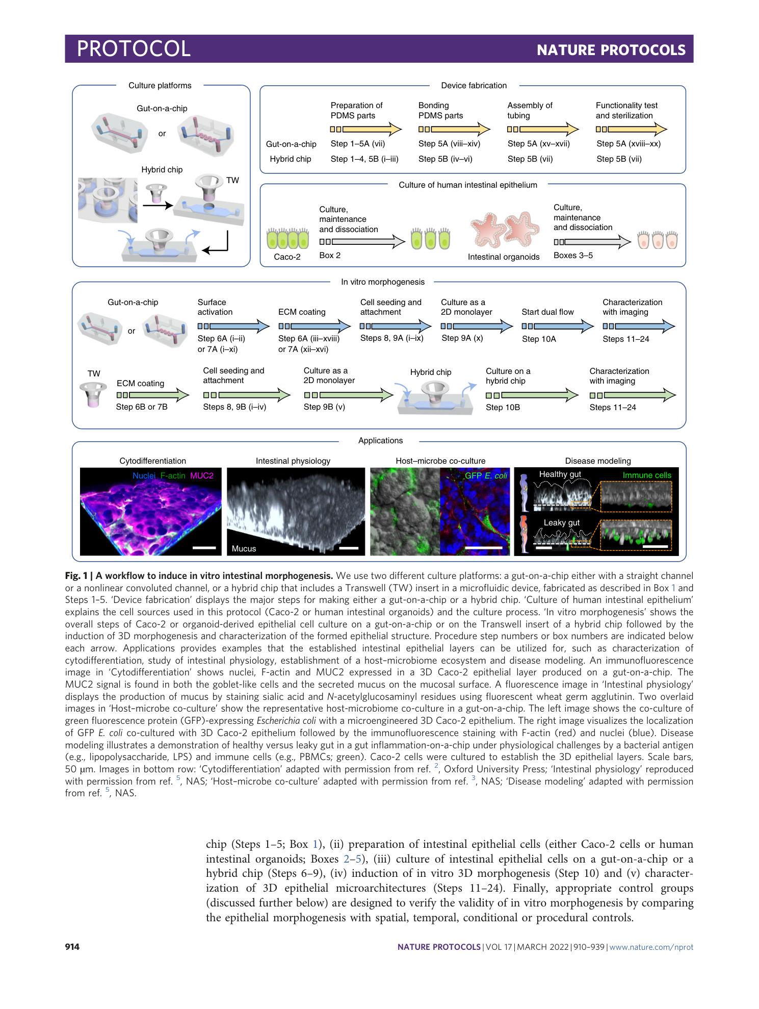
3D in vitro morphogenesis of human intestinal epithelium in a gut-on-a-chip or a hybrid chip with a cell culture insert
Woojung Shin, Hyun Jung Kim
Extended
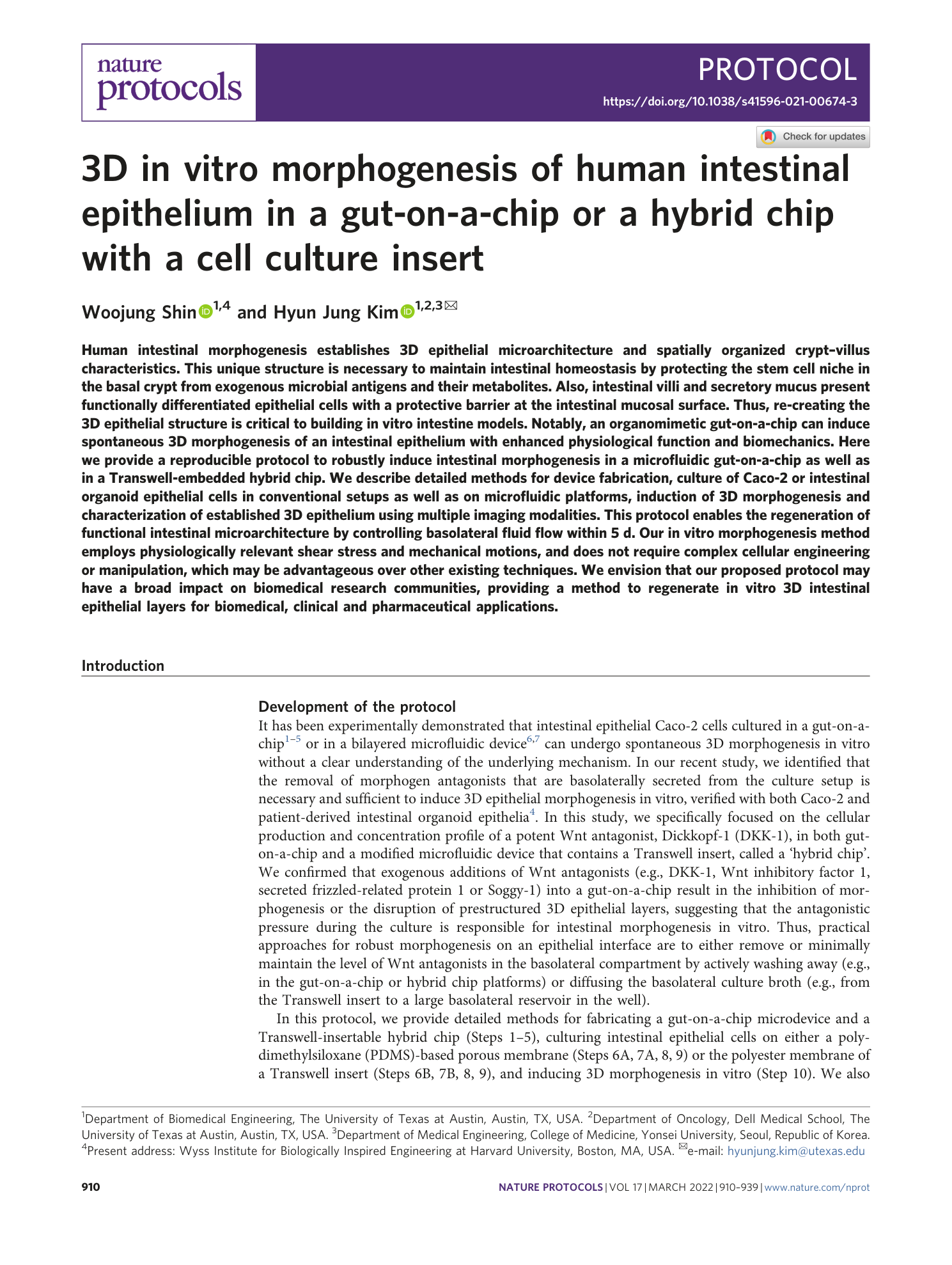
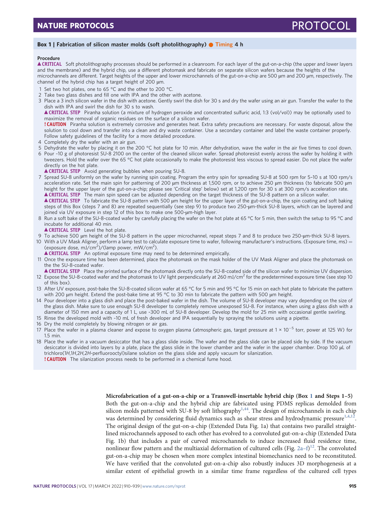
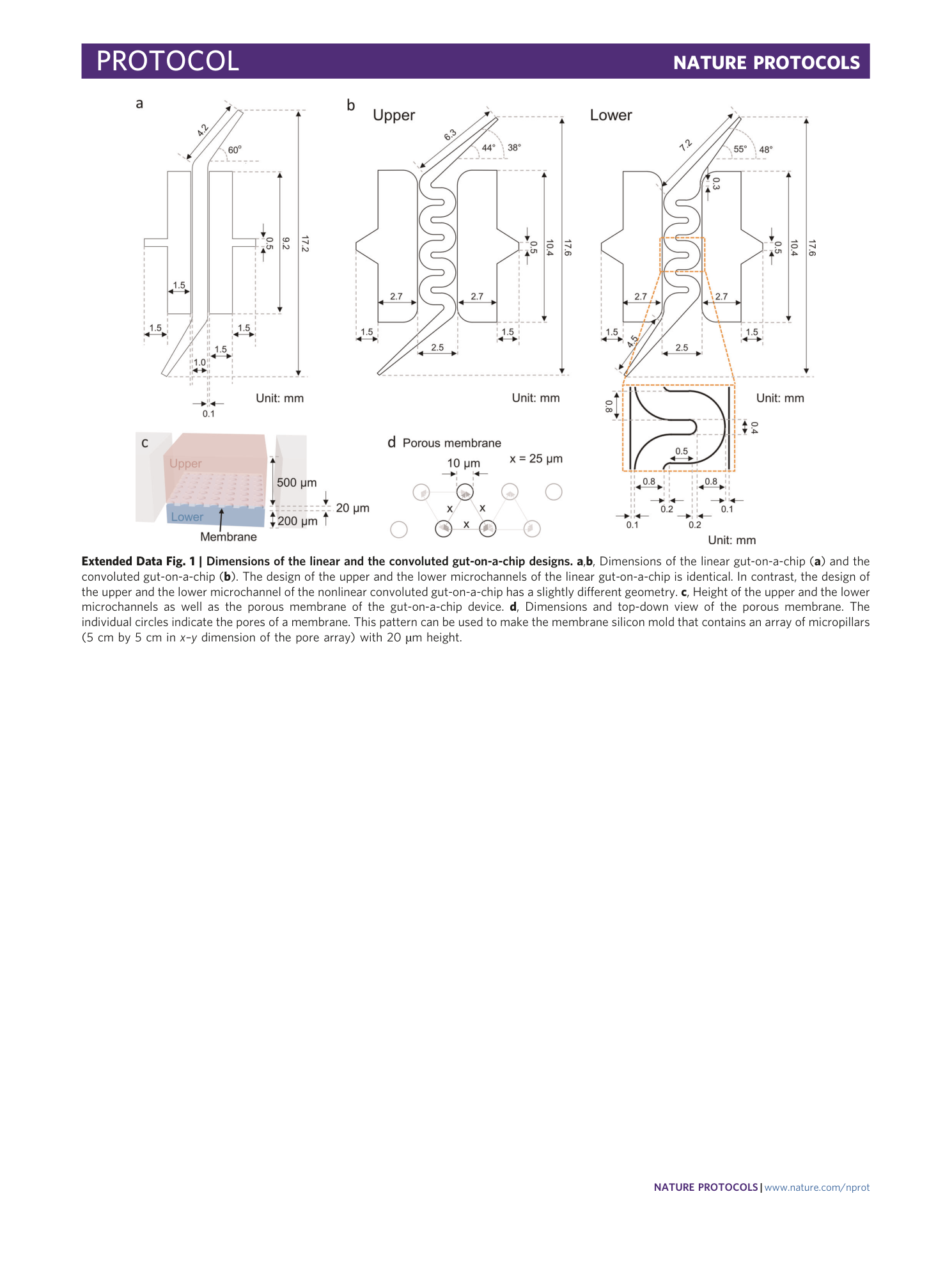
Extended Data Fig. 1 Dimensions of the linear and the convoluted gut-on-a-chip designs.
a , b , Dimensions of the linear gut-on-a-chip ( a ) and the convoluted gut-on-a-chip ( b ). The design of the upper and the lower microchannels of the linear gut-on-a-chip is identical. In contrast, the design of the upper and the lower microchannel of the nonlinear convoluted gut-on-a-chip has a slightly different geometry. c , Height of the upper and the lower microchannels as well as the porous membrane of the gut-on-a-chip device. d , Dimensions and top-down view of the porous membrane. The individual circles indicate the pores of a membrane. This pattern can be used to make the membrane silicon mold that contains an array of micropillars (5 cm by 5 cm in x – y dimension of the pore array) with 20 µm height.
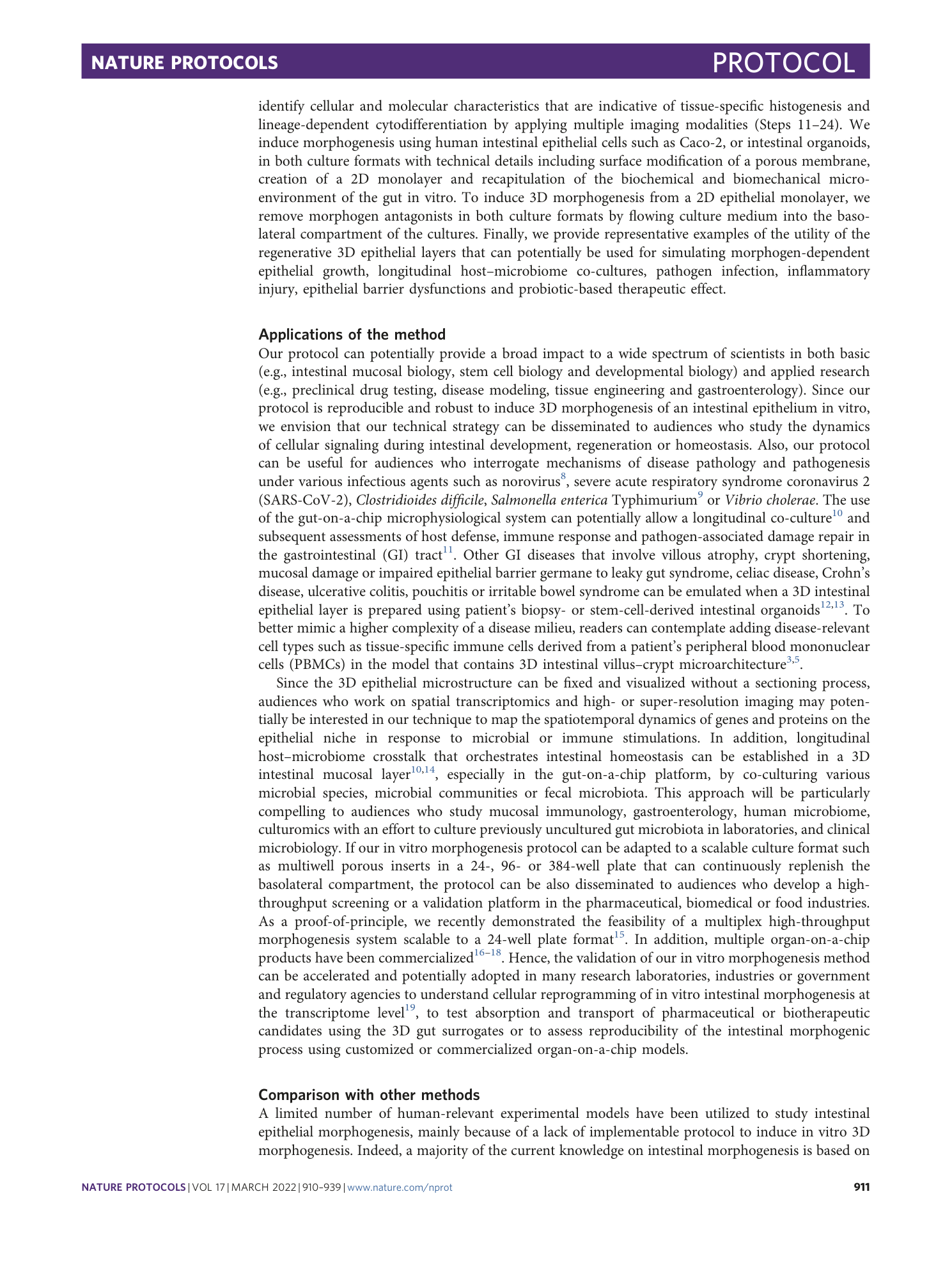
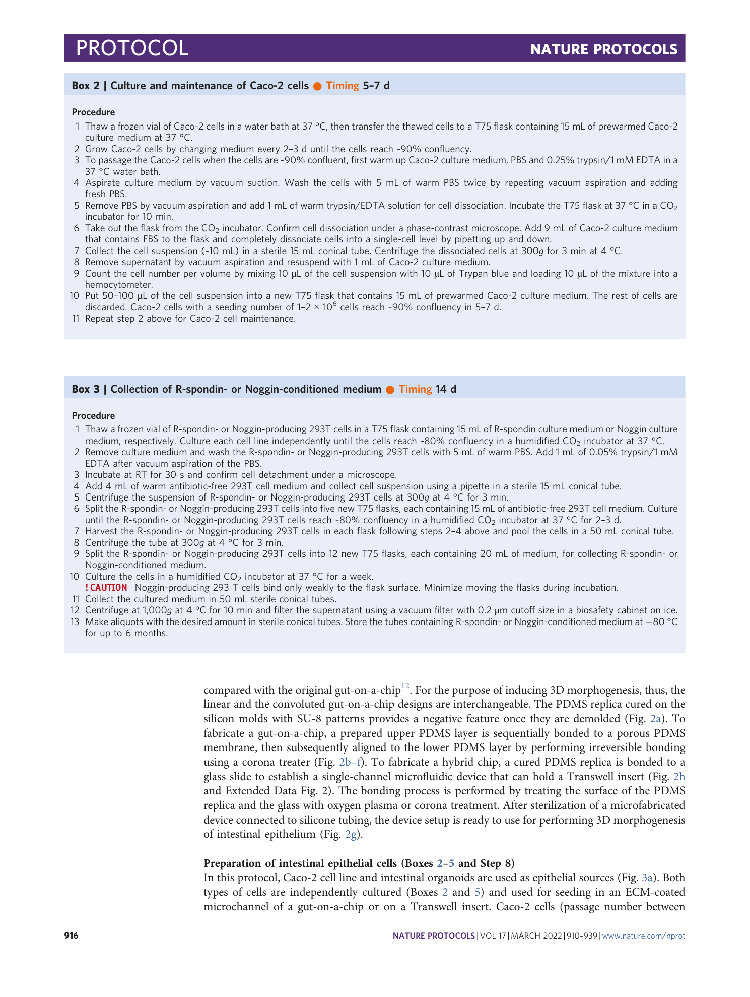
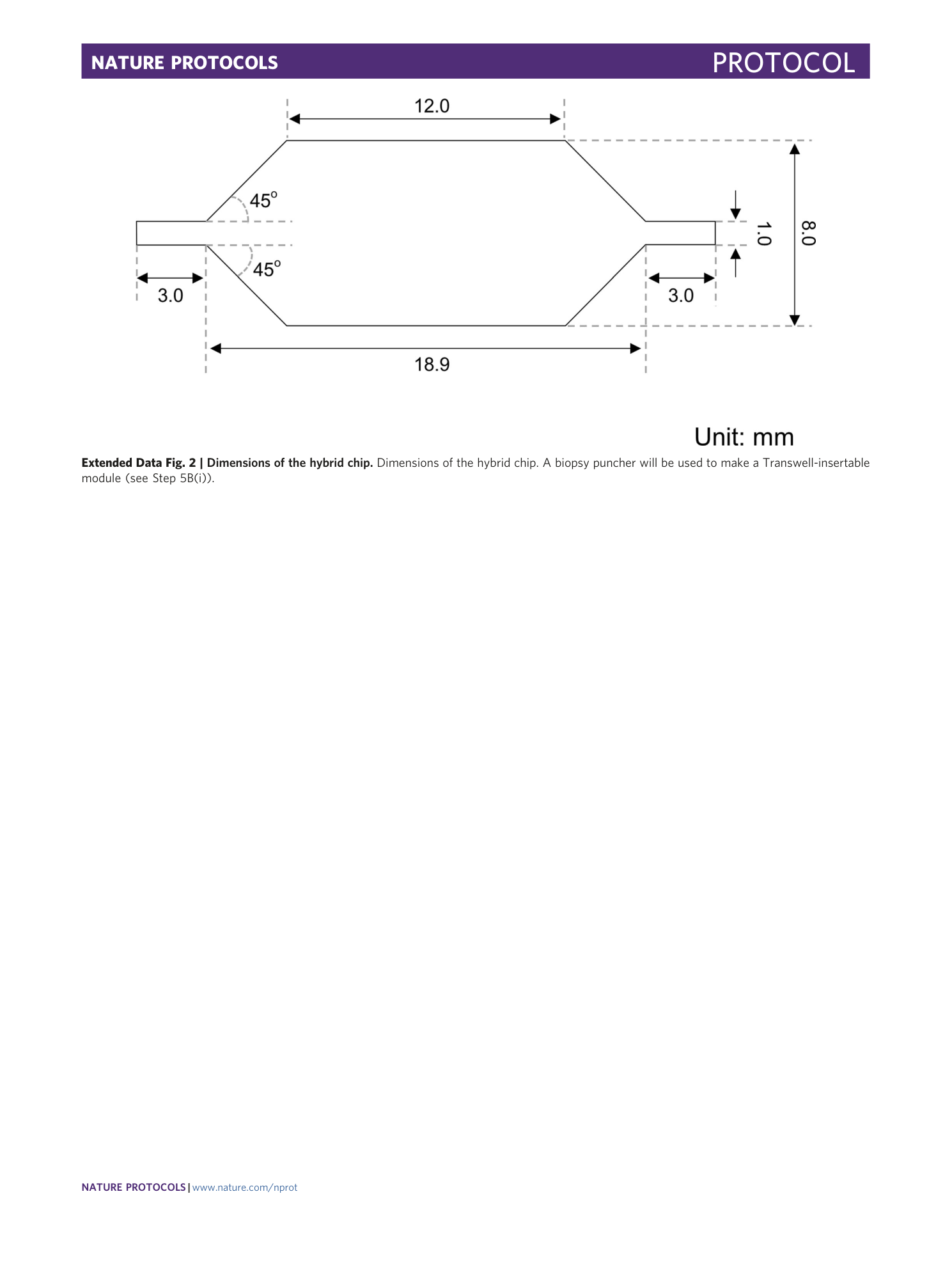
Extended Data Fig. 2 Dimensions of the hybrid chip.
Dimensions of the hybrid chip. A biopsy puncher will be used to make a Transwell-insertable module (see Step 5B(i)).
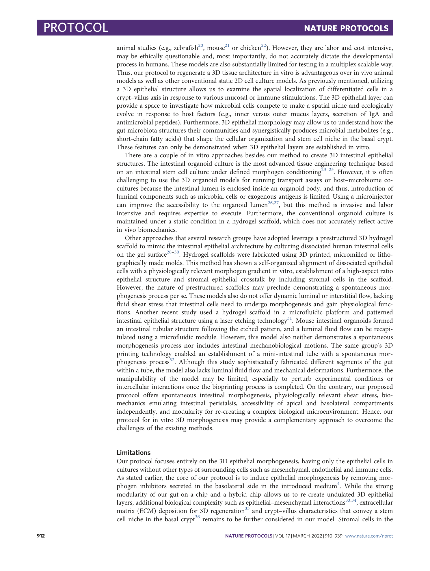
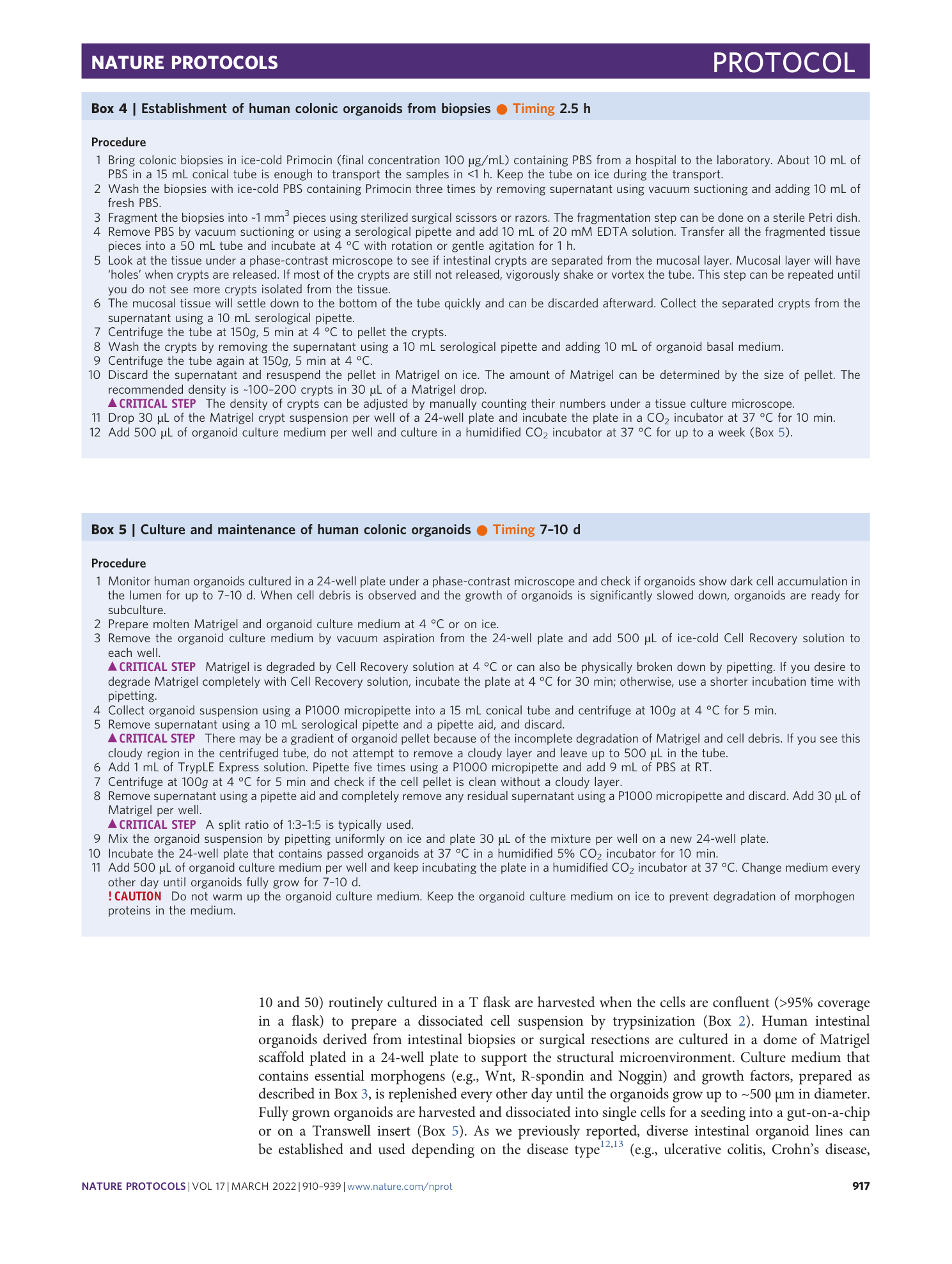
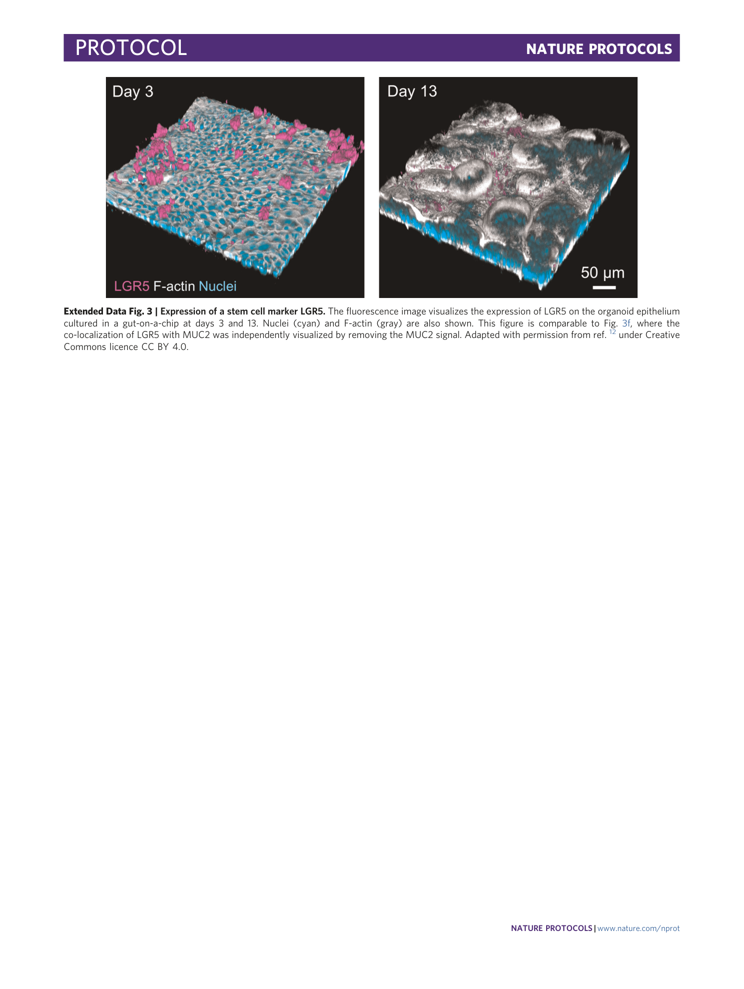
Extended Data Fig. 3 Expression of a stem cell marker LGR5.
The fluorescence image visualizes the expression of LGR5 on the organoid epithelium cultured in a gut-on-a-chip at days 3 and 13. Nuclei (cyan) and F-actin (gray) are also shown. This figure is comparable to Fig. 3f , where the co-localization of LGR5 with MUC2 was independently visualized by removing the MUC2 signal. Adapted with permission from ref. 12 under Creative Commons licence CC BY 4.0.
