Reproducible strategy for excisional skin-wound-healing studies in mice
Matan Yampolsky, Ido Bachelet, Yaron Fuchs


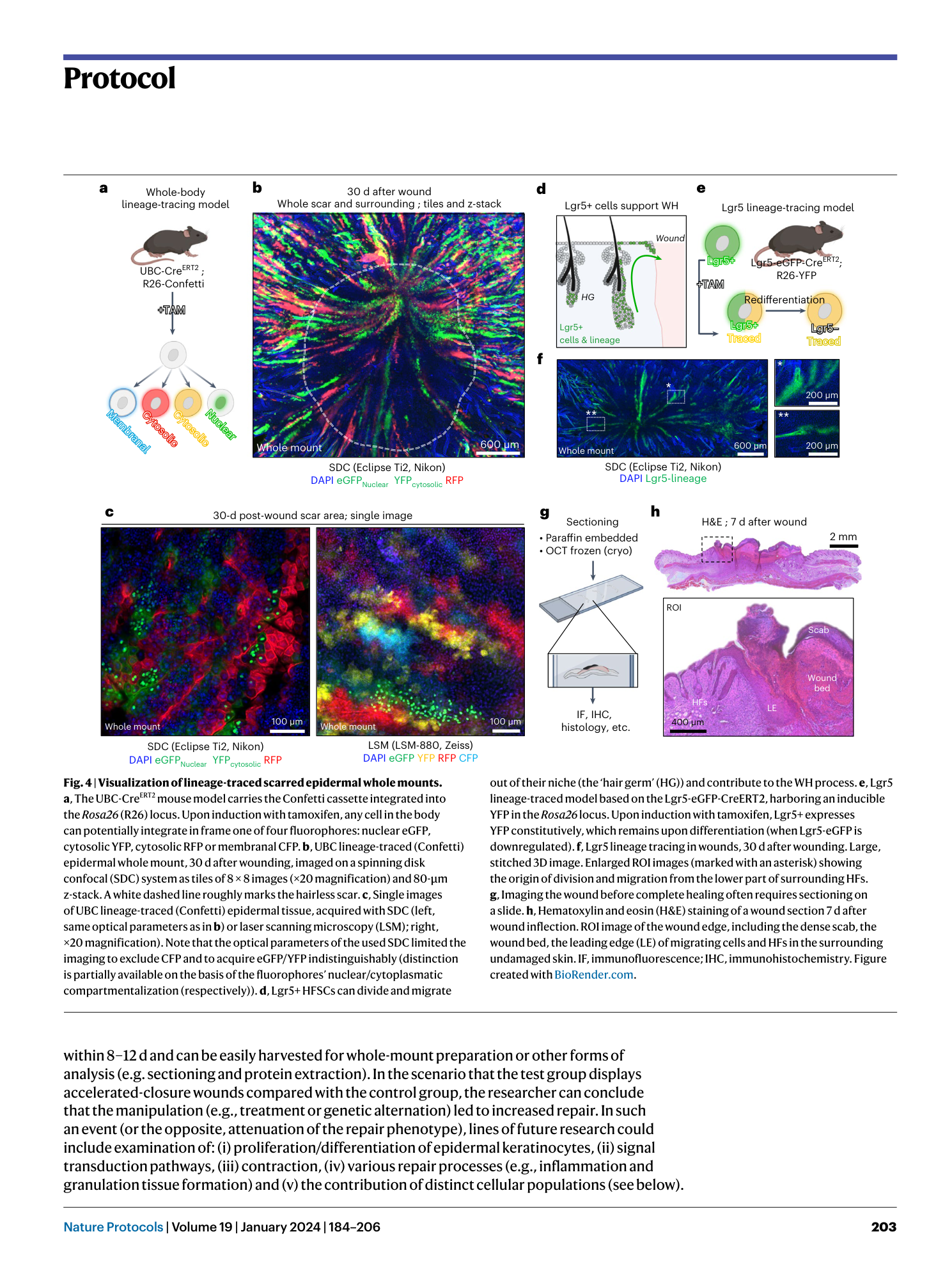
Extended
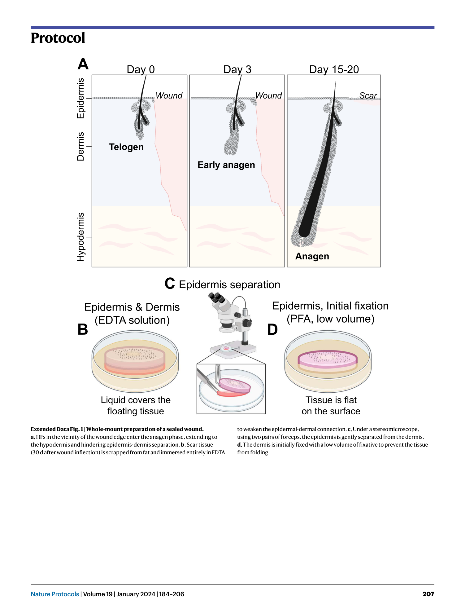
Extended Data Fig. 1 Whole-mount preparation of a sealed wound.
a , HFs in the vicinity of the wound edge enter the anagen phase, extending to the hypodermis and hindering epidermis-dermis separation. b , Scar tissue (30 d after wound inflection) is scrapped from fat and immersed entirely in EDTA to weaken the epidermal-dermal connection. c , Under a stereomicroscope, using two pairs of forceps, the epidermis is gently separated from the dermis. d , The dermis is initially fixed with a low volume of fixative to prevent the tissue from folding.
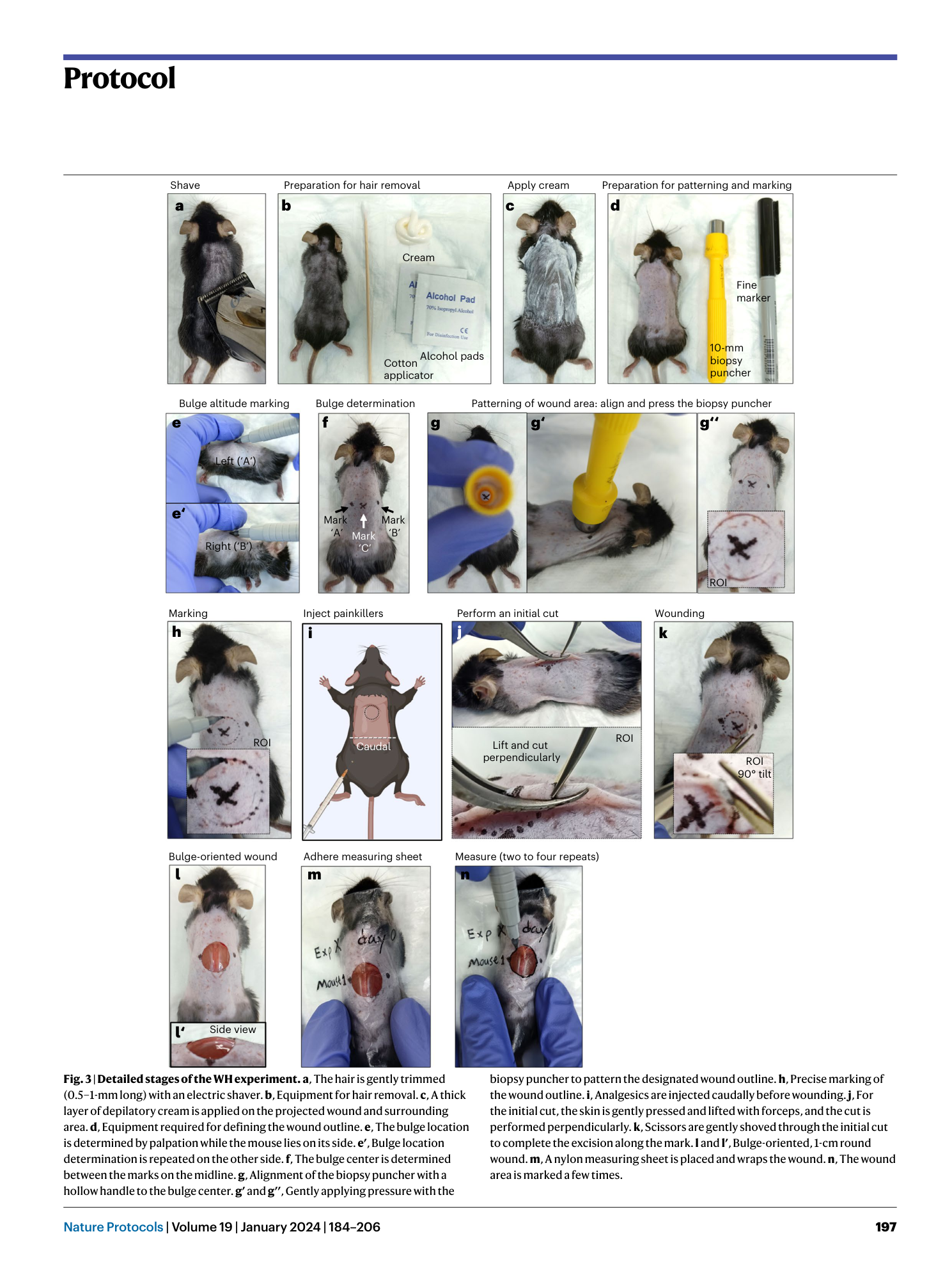
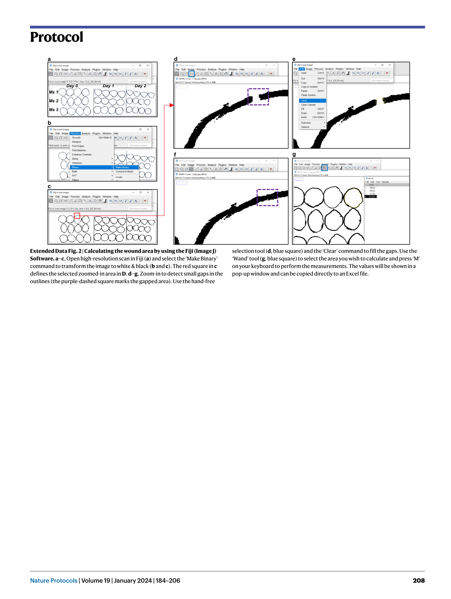
Extended Data Fig. 2 Calculating the wound area by using the Fiji (Image J) Software.
a–c , Open high-resolution scan in Fiji ( a ) and select the ‘Make Binary’ command to transform the image to white & black ( b and c ). The red square in c defines the selected zoomed-in area in D . d–g , Zoom-in to detect small gaps in the outlines (the purple-dashed square marks the gapped area). Use the hand-free selection tool ( d , blue square) and the ‘Clear’ command to fill the gaps. Use the ‘Wand’ tool ( g , blue square) to select the area you wish to calculate and press ‘M’ on your keyboard to perform the measurements. The values will be shown in a pop-up window and can be copied directly to an Excel file.
Supplementary information
Supplementary Information
Supplementary Protocols 1–3, Discussion and Figs. 1 and 2
Supplementary Data 1
Theoretical data analysis template
Supplementary Data 2
Source data for Supplementary Fig. 1B (graph)
