Protocol for infection of Hymenochirus boettgeri with Batrachochytrium dendrobatidis (Bd) and Ranavirus (Rv)
Timothy Y. James, Tamilie Carvalho, Evelyn Faust, Francisco De Jesus Andino, Jacques Robert
Abstract
This protocol outlines the procedures for exposing H. boettgeri specimens to the pathogenic chytrid fungus Batrachochytrium dendrobatidis (Bd) or the Ranavirus Frog Virus 3 (Rv FV3), or both, for co-infection studies. These experiments are necessary to better understand the impacts of these lethal diseases on wildlife and the potential outcome of their interactions on hosts in a controlled laboratory setting. To ensure the safety and accuracy of the experimental exposures, the protocol provides step-by-step instructions and guidelines for handling these pathogens and administering the exposures. All procedures described here were performed in a temperature-controlled room set at 20°C and 13:11 day-night cycle.
Steps
Production of pathogens
Preparation of Bd inoculum
Inoculate several 10 cm Petri dishes containing 1% tryptone media with Bd liquid culture by adding 500µL with a 1000 pipetman. Usually, one plate per 2 frogs is sufficient. Seal all Petri dishes with parafilm.
Inoculate Bd onto new plates from either an actively growing liquid or Petri dish culture to start an inoculum preparation. For liquid inoculation, add ~100µL of an active growing culture and finish by adding 300µL of sterile water. For inoculum from Petri dish cultures, cut 1 cm x 1cm square containing Bd with a lab sterile spatula, then gently rub this onto a new 1% tryptone Petri dish, and finish by adding 300µL of sterile water. Seal all Petri dishes with parafilm.
Keep plates at 20°C and check daily for maximum zoospore production.
Add 2mL of sterile water to the Bd culture plates. Wait for 1h 0m 0s, then gently collect the water containing Bd zoospores by pipetting.
Transfer all the liquid into a 50 mL sterile Falcon tube. If any clumps of zoosporangia are noted, filter the inoculum using a 40 μL cell strainer (431750).
Calculate zoospore concentration using either a hemocytometer or Coulter counter. If necessary, dilute by adding sterile water.
All procedures describe here should be conducted in a sterile condition, ideally in a biological safety cabinet.
Cell line and FV3 high titer stocks production
Maintain baby hamster kidney-21 cells (BHK-21; ATCC no. CCL-10) in Dulbecco's modified Eagle's medium supplemented with 10% fetal bovine serum, penicillin (100 U/mL) and streptomycin (100 µg/mL), in a sterile cell culture flask with a vented cap, with 5% CO2 at 37°C.
Produce FV3 (Granoff et al., 1965; ATCCVR-569 strain) stocks by a single infection passage on BHK-21 cells, culture for 168h 0m 0s at 30°C in 5% CO2 (in 250mL cell culture vented flasks).
Purify FV3 virus particles by ultracentrifugation on a 30% sucrose gradient and quantification by plaque assay on BHK-21 monolayers in 6-well plates under an overlay of 1% methylcellulose (Morales et al., 2010).
Aspirate the overlay media and stain the cells for 10 min with 1% crystal violet in 20% ethanol before counting.
FV3 or Bd Exposure
Water Bath for FV3 and/or Bd
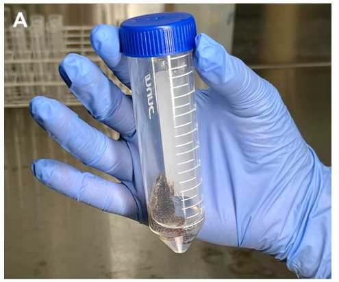
Begin by adding at least 3 mL of conditioned water, as described in the “Housing and Care for Hymenochirus boettgeri”, containing the desired concentration of FV3, Bd, or both, to a 50 mL Falcon tube (Figure 1A).

Change sterile gloves for each animal handled. Place an individual adult amphibian into each Falcon tube and keep them in the water bath for 2-3 hours.
After this exposure period, transfer the animals back to their permanent tanks.
Buccal Inoculation for FV3 Exposure
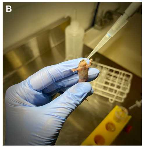
With sterile gloves for each animal, hold the amphibian by its legs with one finger supporting its back and head (Figure 1B).

Open its mouth by gently inserting and twisting a sterilized guitar pick, or similar device, in the side of the mouth opening. Using a pipetman and 100 μL pipette tips, slowly inject up to 40 μL of the desired concentration of FV3 inoculum into the amphibian's mouth.
Keep the amphibian still with the pipette tip inside its mouth for 5 seconds to prevent regurgitation.
Place the animal back in its permanent tank and repeat the procedure, if necessary to achieve total exposure dose, after at least 30 minutes to minimize stress on the animal.
FV3 Exposure by Intraperitoneal Injection
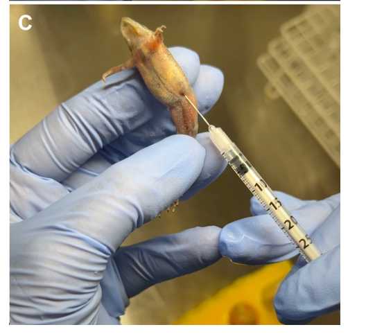
Using sterile gloves for each animal, hold the amphibian by its legs with its belly exposed and turned upward, supporting its back (Figure 1C). The frog should be immobilized. We sterilize the area of injection using 75% EtOH first, then inject FV3.

Carefully insert a needle at a shallow angle just anterior to and above the thigh, aiming toward the head (45-degree angle).
Gently push the syringe until you feel a slight 'pop,' indicating the needle is inside the peritoneal cavity, and then slowly release the liquid, injecting up to 60 μL. Place the animal back in its permanent tank.
Dose responses
We have observed mortality of H. boettgeri adults given the following dose ranges: 104 to 107 Bd zoospores per mL (varying with Bd strain), 106 to 107 FV3 plaque forming units (PFU) per mL in water bath exposure, 106 to 107 total PFUs in buccal inoculations, and 104 to 106 total PFUs in intraperitoneal injections.
Overall, for both pathogens and all exposure methods, higher doses lead to faster animal mortality and reduced chances of recovery from infection.
However, we acknowledge that susceptibility may vary not only with the dose but also with pathogen genotype and host characteristics, such as microbiome composition and prior infections. Therefore, we recommend conducting a pilot study to determine the optimal dose for both pathogens.

