Juxtacellular Recordings in Ventral tegmental area and Locus coeruleus
Matthias Prigge, Cristian González-Cabrera, Kaushik S. More
Abstract
This protocol describes juxtacellular recordings in the VTA and LC of adult mice, involving anesthesia, precise brain targeting, and neuron labeling. Using specialized electrodes and a dual-channel amplifier, neuronal activity is recorded and modulated with current injections. Following recordings, brains are perfused, fixed, and sectioned for detailed morphological analysis.
Steps
Preparation of Adult Mice for juxtacellular recordings
Use adult mice weighing 25-30 g.* Anesthetize the animal with urethane at a dosage of 1.25 g/kg.
-
Place the mouse in the stereotaxic frame and align the head angle according to Bregma and Lambda, with a tolerance of a maximum of 50 microns between them.
-
Expose the skull by shaving the mouse's head and making a sagittal incision on the skin.
-
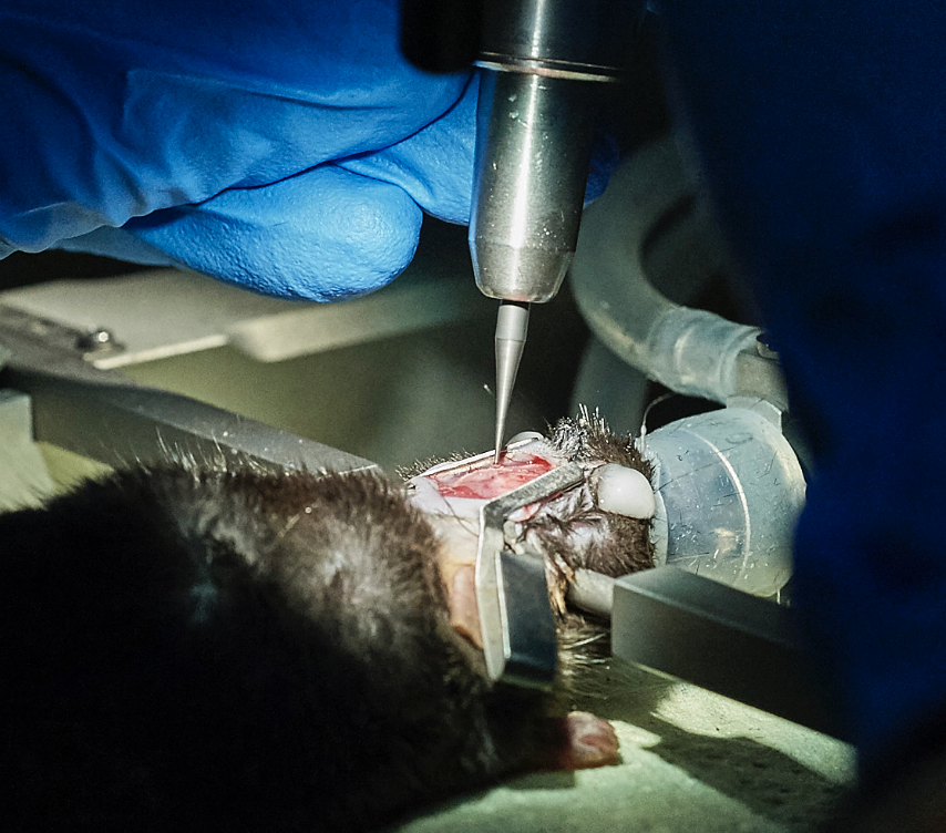
Find the coordinates and drill the bone. Once the dura is exposed, carefully remove it with the forceps (n°).
VTA: A/P 3.25, L/M 0.5
LC: A/P 5.34, L/M 1.1
- Drill a small hole around Lambda and install the reference screw.
Juxtacellular set-up
Employ an intracellular recording amplifier (NeuroData Dual channel IR-283), an analog-to-digital converter (CED Micro-1401-3), and the Spike2 software..* Use 5-16 MΩ (40-45 MΩ for intracellular) glass electrodes with a tip diameter of less than 1 μm.
- Fill the electrodes with a solution containing:
- 250 mM k-gluconate, 5 mM KCl, 1 mM MgCl2, 2 mM EGTA, 5 mM HEPES, 2 mM MgATP, and Tetramethyl-Rhodamin Biocytin tracer or Neurobiotin (2% w/v); pH 7.2.
Juxtacellular/Intracellular Recordings and Labelling
After penetrating the brain, ensure the electrode impedance is correct by injecting 0.5 nA pulses and adjusting the bridge/balance module.* Move down the electrode at a constant low speed until reaching the dorsal VTA (z=-4.0).
- Start the exploration by moving down not faster than a micron per second.
- Isolate spontaneously active neurons until the extracellular spike amplitude is approximately 0.7 to 1.0 mV (for intracellular penetration about 2.0-2.5 mV.)
- After baseline recordings, label neurons using the juxtacellular (Pinault, 1996) or intracellular methods.
For juxtacellular labeling:* Label neurons by injecting a small current (1-4 nA for 5-10 minutes, 250 ms ON-250 ms OFF). Make sure that the neuron is being modulated by the current injection.
For intracellular labeling:* Apply AC pulses to gain intracellular access. If unsuccessful, adjust by 2 micrometers and reapply AC pulses.
- Once intracellular access is achieved, stabilize neurons with negative current.
- Label neurons by injecting a small current (1-2 nA for 10-15 minutes, 250 ms ON-250 ms OFF).
Wait for at least 2 hours before perfusion.
Post-Recording and Labeling Process:
Perfuse the animals via the ascending aorta with 0.01 M PBS followed by 4% paraformaldehyde in PBS.* Post-fix brains between 6-12 hours in 4% PFA.
- Cryoprotect the brain in 30% sucrose until it sinks.
- Section the brains at 40 μm using a freezing microtome.
