Intravital microscopy of dynamic single-cell behavior in mouse mammary tissue
Jane E. Visvader, Anne C. Rios, Caleb A. Dawson, Scott N. Mueller, Geoffrey J. Lindeman
Intravital microscopy
Mouse mammary tissue
Single-cell behavior
Multiphoton imaging
3D video processing
Supplementary information
Supplementary Information
Supplementary Discussion.
Reporting Summary
Supplementary Video 1
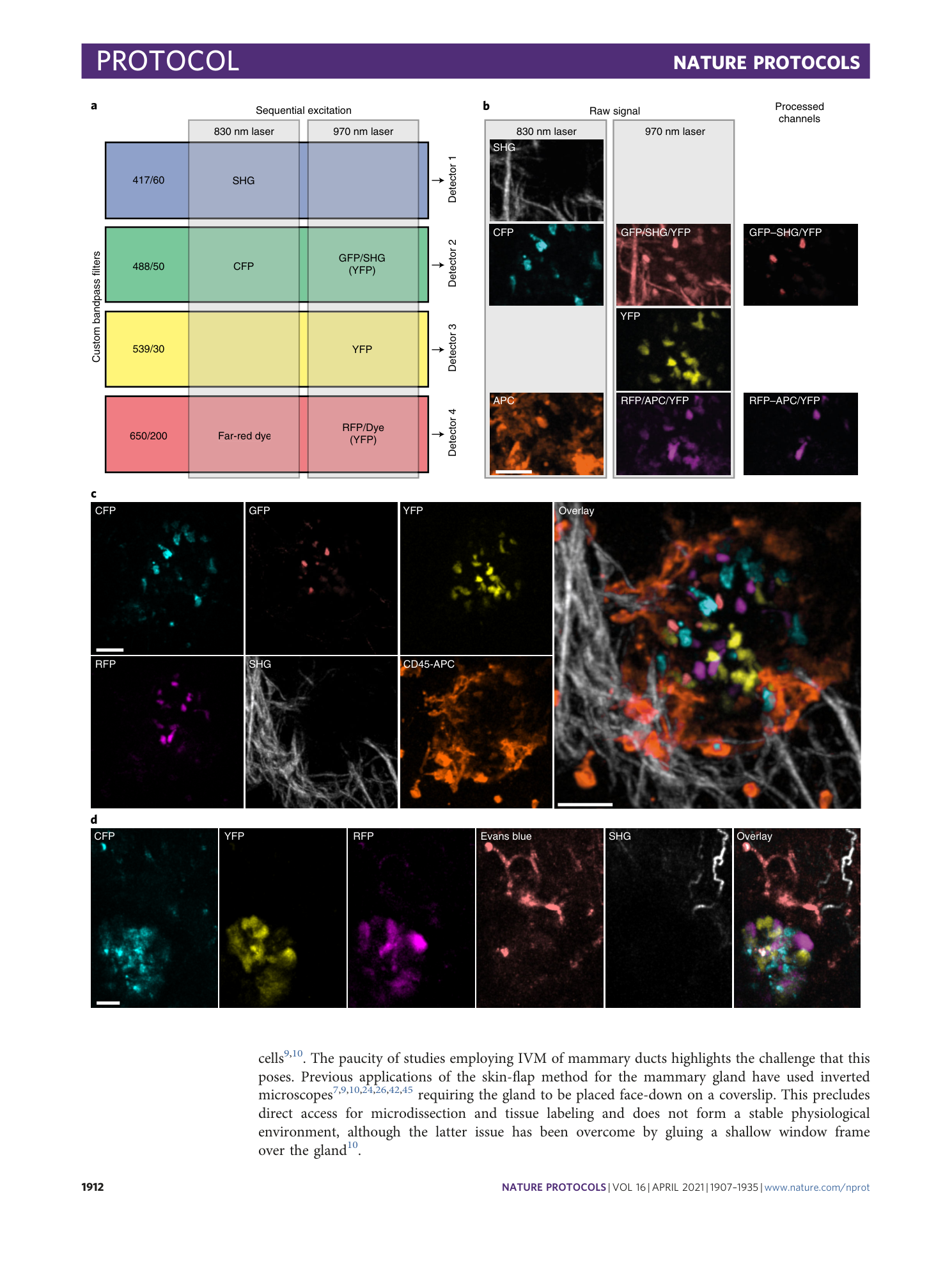
Six-color IVM of immune cell interactions with mammary terminal end bud cap cells during morphogenesis in puberty . IVM of a TEB in a 5 week-old K5/TetCre/Confetti mouse treated with doxycycline for 3 d at 4 weeks of age and labeled with CD45 APC antibody placed in the imaging chamber ( n = 2 mice). CFP, cyan; GFP, pink; YFP, yellow; RFP, magenta; SHG, gray; CD45, orange. Related to Fig. 3c. All experiments were approved by the Walter and Eliza Hall Institute Animal Ethics Committee.
Supplementary Video 2
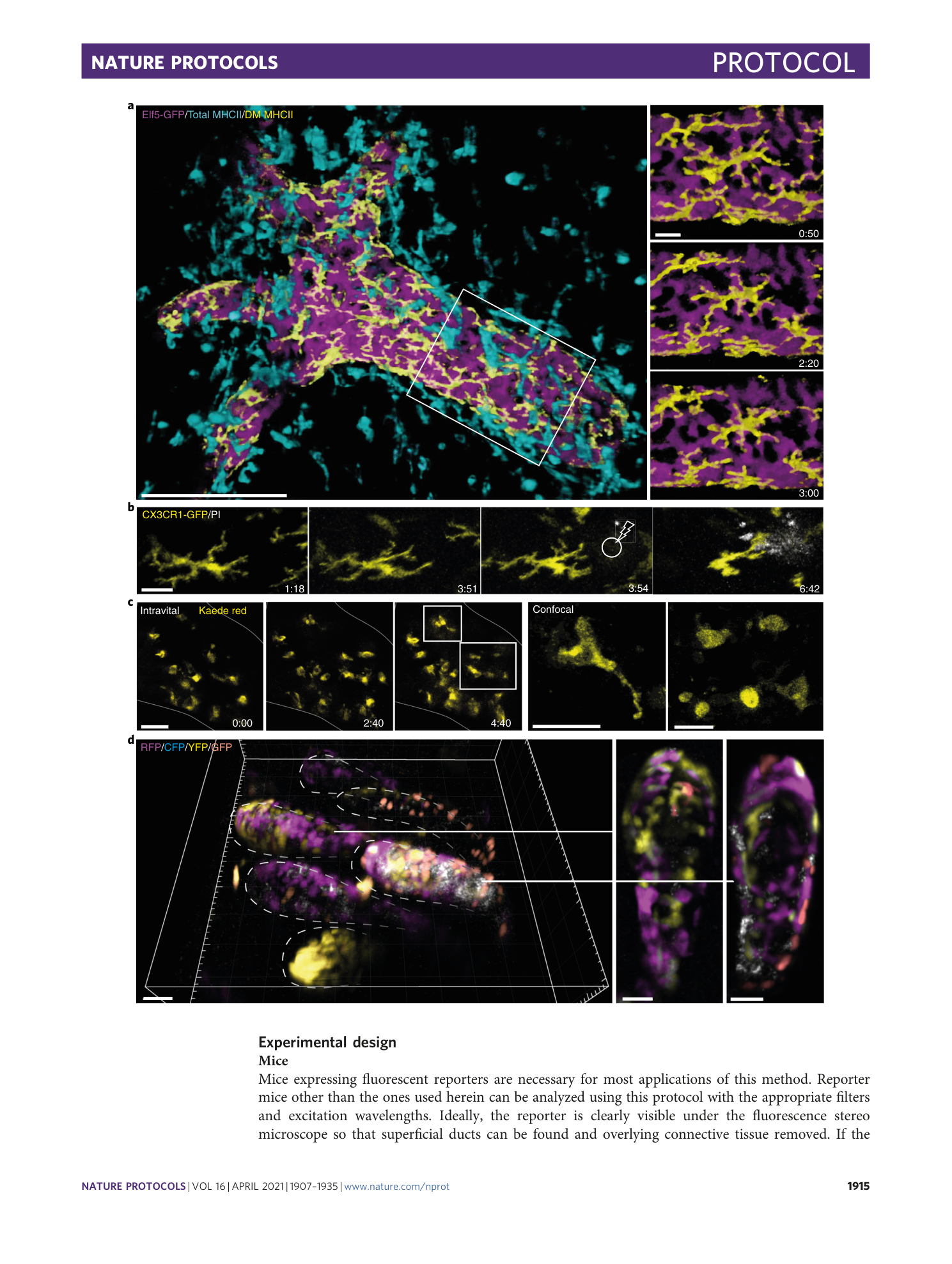
IVM of resident ductal macrophage behavior in mammary ducts . Animated IVM of mammary ducts in an Elf5-GFP mouse with immunolabeling by MHCII Alexa Fluor 647 antibody in the imaging chamber 12 . The movie cycles over a 6 h time span with images acquired every 10 min. GFP, magenta; Duct-adjacent MHCII, yellow; stromal MHCII, cyan; collagen SHG, pink. Duct-adjacent MHCII was isolated in Imaris by creating a low resolution GFP surface and masking the MHCII signal. The duct structure is 300 µm across ( n = 6 mice). Related to Figure 5a. All experiments were approved by the Walter and Eliza Hall Institute Animal Ethics Committee.
Supplementary Video 3
IVM of the resident macrophage response to damage of the mammary epithelium . An animation of IVM of a mammary duct in a Cx3cr1 GFP/+ mouse 12 . The movie cycles through time points prior to damage showing the arrangement of GFP-high ductal macrophages (yellow) around a duct, then views an optical section through ductal macrophages before and after precise photoablation at 4 h (bolt symbol). Images were acquired every 3 min ( n = 3 mice). Related to Figure 5b. All experiments were approved by the Walter and Eliza Hall Institute Animal Ethics Committee.
Supplementary Video 4
IVM of single photoactivated mammary terminal end bud cells . IVM of a TEB in a 4-week-old Kaede mouse after photoconversion of single cells using the Olympus SIM scanner ( n = 2 mice). Related to Figure 5c. All experiments were approved by the Walter and Eliza Hall Institute Animal Ethics Committee.
Supplementary Video 5
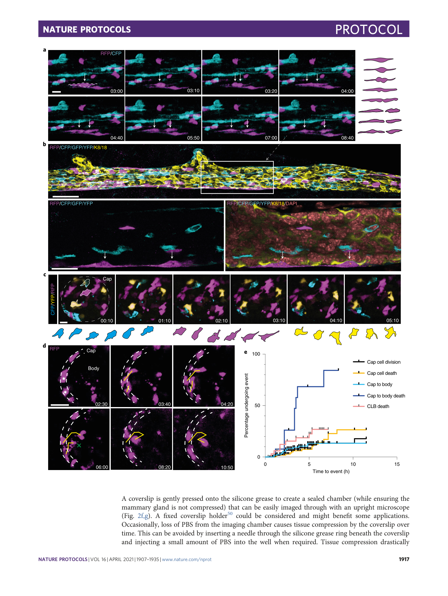
IVM of mammary duct basal cell division . IVM of a duct in an 8-week-old K5/TetCre/Confetti mouse after treatment with doxycycline at 4 weeks of age showing a myoepithelial cell dividing longitudinally ( n = 5 mice). The duct was imaged every 10 min for 8 h and 40 min. Related to Fig. 6a. All experiments were approved by the Walter and Eliza Hall Institute Animal Ethics Committee.
Supplementary Video 6
IVM of mammary duct basal cells, example 2 . IVM of a duct in a 6-week-old K5/TetCre/Confetti mouse after treatment with doxycycline at 4 weeks of age ( n = 5 mice). The duct was imaged every 10 min for 4 h. All experiments were approved by the Walter and Eliza Hall Institute Animal Ethics Committee.
Supplementary Video 7
IVM of mammary terminal end bud cap cell dynamics . IVM of a TEB in a 5-week-old K5/TetCre/Confetti mouse treated with doxycycline for 3 d at 4 weeks of age. An optical slice through the outer cap layer is shown to reveal cap cell dynamics ( n = 4 mice). Related to Fig. 6c. The TEB is pointing upward. All experiments were approved by the Walter and Eliza Hall Institute Animal Ethics Committee.
Supplementary Video 8
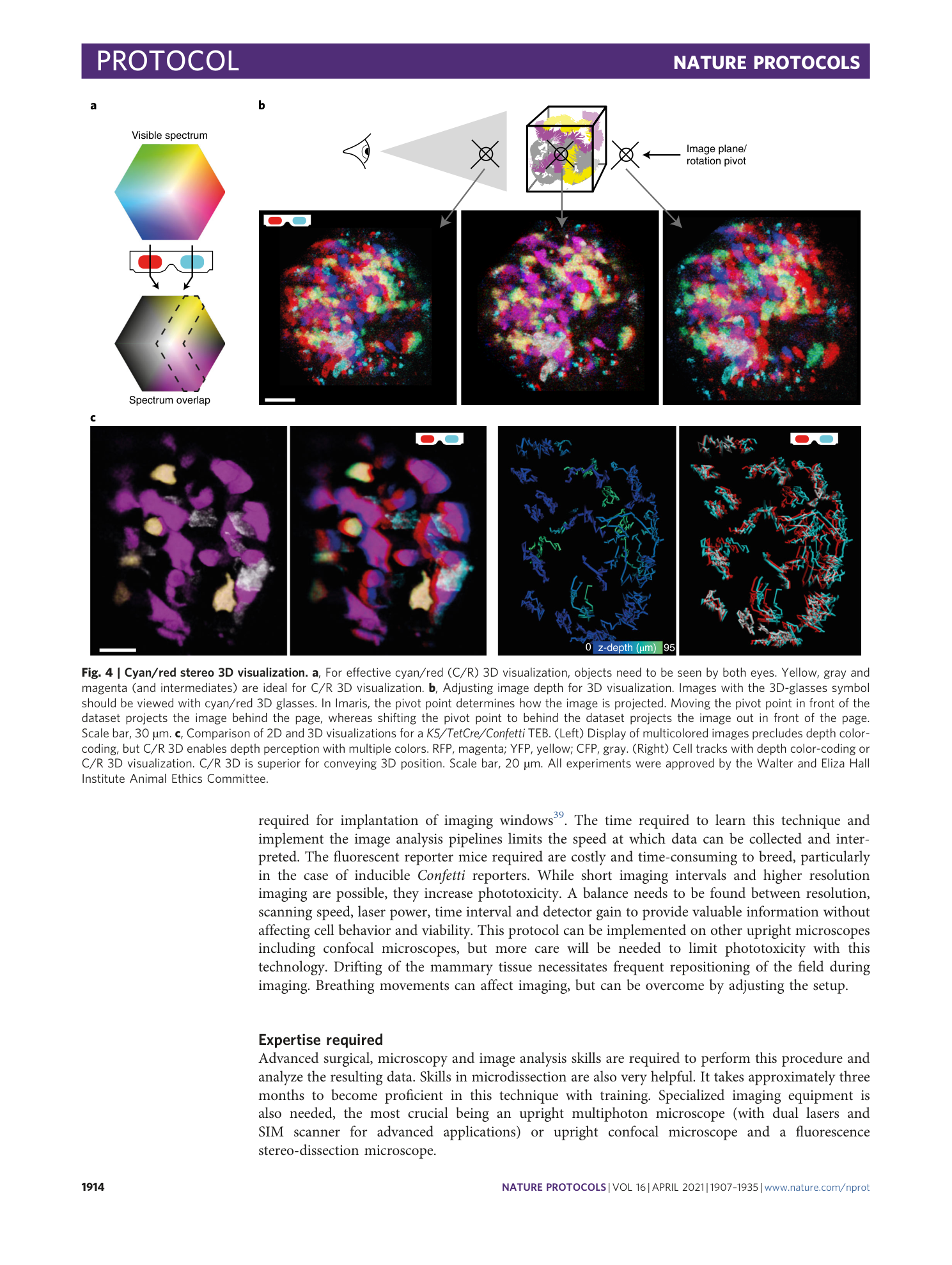
IVM of mammary terminal end bud cap cells in stereo 3D . IVM of a TEB in a 5-week-old K5/TetCre/Confetti mouse treated with doxycycline for 3 d at 4 weeks of age. The full image volume is displayed for viewing with cyan/red 3D glasses. The TEB is pointing to the left ( n = 4 mice). Related to Fig. 6c-d. All experiments were approved by the Walter and Eliza Hall Institute Animal Ethics Committee.
Supplementary Video 9
IVM of mammary cap cell migration into the terminal end bud body . IVM of a TEB in a 5 week-old K5/TetCre/Confetti mouse treated with doxycycline for 3 d at 4 weeks of age. An optical slice through the center of the TEB (which is pointing to the left), showing cap cells that migrate from the cap into the TEB body. RFP, magenta ( n = 4 mice). Related to Fig. 6d. All experiments were approved by the Walter and Eliza Hall Institute Animal Ethics Committee.
Supplementary Video 10
IVM of mammary terminal end bud cap cells, example 2 . IVM of a TEB in a 5 week-old K5/TetCre/Confetti mouse treated with doxycycline for 3 d at 4 weeks of age ( n = 4 mice). The TEB is pointing to the right. All experiments were approved by the Walter and Eliza Hall Institute Animal Ethics Committee.
