Chronic, cortex-wide imaging of specific cell populations during behavior
Joao Couto, Simon Musall, Xiaonan R. Sun, Anup Khanal, Steven Gluf, Shreya Saxena, Ian Kinsella, Taiga Abe, John P. Cunningham, Liam Paninski, Anne K. Churchland


Extended
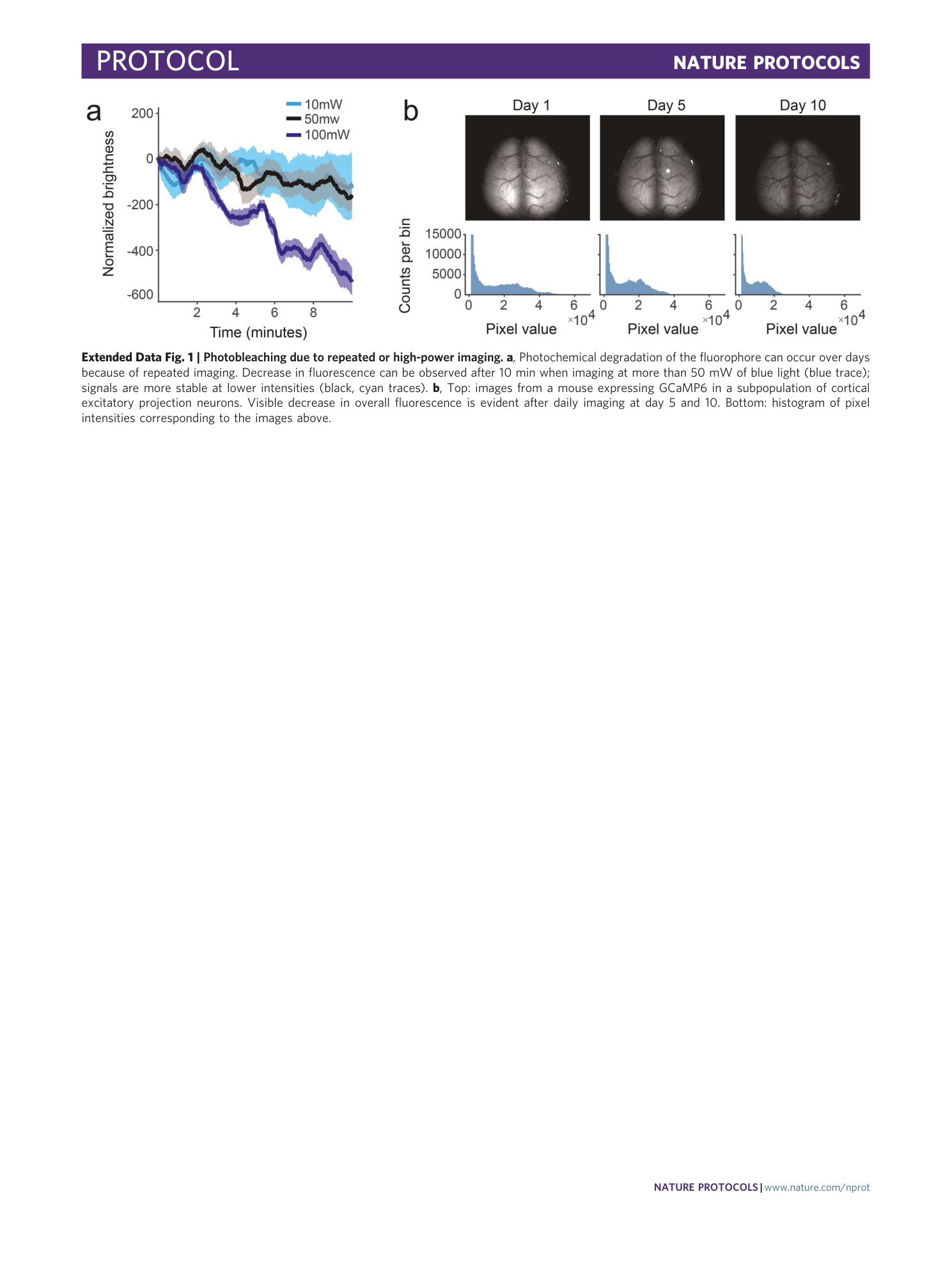
Extended Data Fig. 1 Photobleaching due to repeated or high-power imaging.
a , Photochemical degradation of the fluorophore can occur over days because of repeated imaging. Decrease in fluorescence can be observed after 10 min when imaging at more than 50 mW of blue light (blue trace); signals are more stable at lower intensities (black, cyan traces). b , Top: images from a mouse expressing GCaMP6 in a subpopulation of cortical excitatory projection neurons. Visible decrease in overall fluorescence is evident after daily imaging at day 5 and 10. Bottom: histogram of pixel intensities corresponding to the images above.
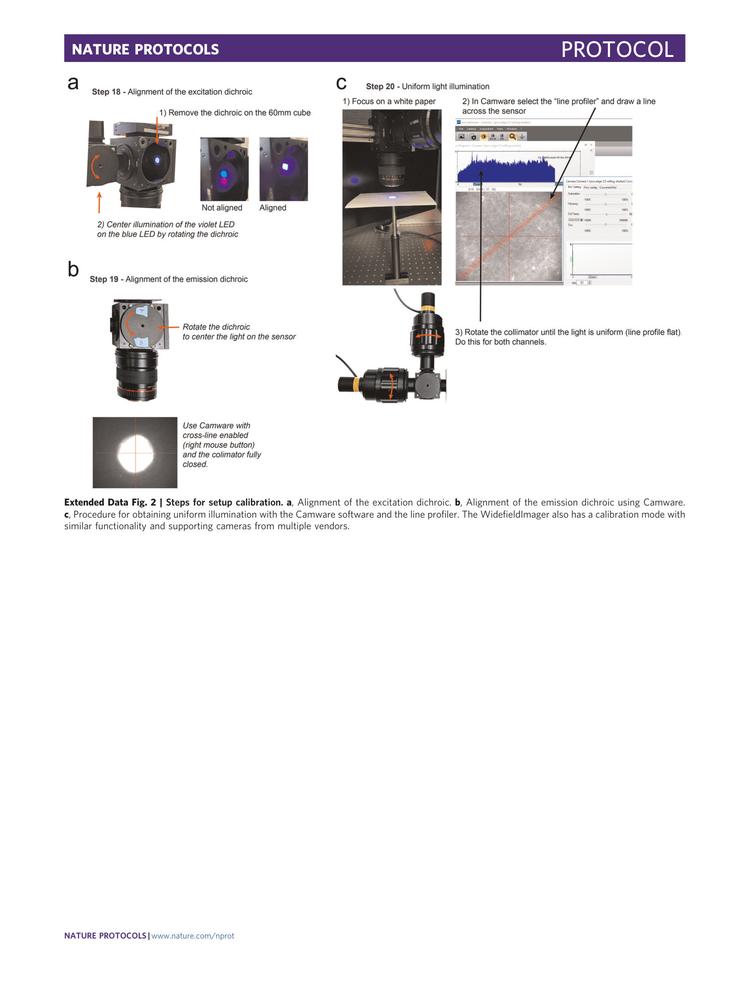
Extended Data Fig. 2 Steps for setup calibration.
a , Alignment of the excitation dichroic. b , Alignment of the emission dichroic using Camware. c , Procedure for obtaining uniform illumination with the Camware software and the line profiler. The WidefieldImager also has a calibration mode with similar functionality and supporting cameras from multiple vendors.
