Light microscopy-based neuron tracing and reconstruction
Yoland Smith, Thomas Wichmann, Rosa Villalba
Abstract
This protocol details the term ‘neuron reconstruction’ is used here to refer to the process of delineating the different components of neurons (soma, axon, dendrites, and dendritic spines) using light microscopy, to create a geometric model of these cells for subsequent quantitative analyses of aspects of their morphology.
Steps
Step by step protocol (based on Neurolucida system):
Log in/switch on the computer, stage controller and joystick, microscope lamp and camera.
Start observations at the objective lens with the lowest magnification, e.g., 2.5x.
Place and secure the slide on the microscope stage.
Using the joystick, move the microscope stage to center the region of interest (ROI) of the tissue under the objective lens (XY movements).
Once the ROI is centered under the objective:
Open the software (Neurolucida). On the toolbar select Acquire → Live Image.
A live image of the field of view will be shown on the computer screen.
Using the microscope binoculars, focus on the ROI using different objective lenses (as needed) and select the neuron to be traced (see also below).
Very important:
- There are two types of objective lenses which are not interchangeable.
- Dry objectives: 2.5x, 5x, 10x, 20x
- Immersion oil objectives: 40x, 100x
- Design the system to be used with the microscope's fine focus knob (usually on the side of the joystick box). Slowly turn the knob when adjusting the focus.
Start with the low magnification objective (2.5x) to find the ROI and gradually increase the microscope’s magnification (5x→10x→20x) to identify the neuron selected to be traced.
While you increase the magnification, adjust the focus and brightness of your image.
Adjust the overall brightness of the image by using the Lamp Power Box. Additionally, tools provided by the Neurolucida software can help to optimize the dynamic range and signal-to-noise ratio of the image.
- Check the Camera Histogram and adjust the brightness until the grey level reaches the end of the histogram.
- Under ‘Camera Settings ’, adjust exposure and gain to improve image quality.

Before using the immersion oil objectives (40x, 100x) add one drop of the immersion oil to the microscope slide (on top of the ROI).
For neuron tracing, use the 100x immersion oil objective.
Very important:
- Double-check to ensure that Neurolucida is set to “100x–oil”
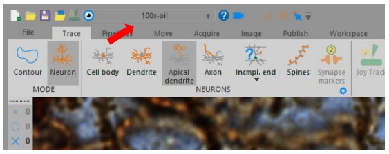
Neurolucida allows tracing of many tissue elements. The following is an example on the procedures used to trace the outline of a neuron, its dendrites, and dendritic spines.
Under ‘Trace’, select Neuron→Cell body.
Adjust the focus to the cell body of the neuron to be traced. Click on the cell body in the image to create a reference point.
Once the cell body is traced, finish the tracing by right-clicking and select “Finish Cell Body”. Once complete, save and label the file appropriately, using a consistent naming scheme across all samples of the study (e.g.,AnimalID_Slice##_BrainRegion_Neuron##). Add an identifier of the side of the hemisphere, as needed.
In the Structure Selection list, choose the next structure to be traced (i.e., dendrite/basilar dendrites, apical dendrite). Before starting to trace dendrites, set the position of the Z meter to “zero” by simultaneously holding the ALT and S keys.
- As you trace dendrites,
- Adjust the focus. Neurolucida keeps track of the Z-depth of the tracings.
- Use the mouse wheel to set and/or adjust the circular cursor to match the diameter of the process.
- When tracing a dendrite, bifurcating junction (i.e., branches) may be encountered. If this happens, right-click and select “Bifurcation” and a “node” marker will be placed. Continue tracing the “main” branch. Once you get to the end of the branch, right-click and select “Finish Ending”. The Neurolucida software will automatically jump back to the previously identified bifurcating nodes so that remaining branches can be traced.
After all dendrites and their branches have been traced, the user can manually mark other relevant tissue elements. For example, to mark the total number of dendritic spines, select the “Spine Tool” and then “None” for dendritic spine shape. While following the traces of the dendrite/s and the different branches adjust the focus to be able to identify and mark the dendritic spines along the dendritic trees.
Once the neuronal reconstruction is complete, morphometric analysis of the neuronal processes and dendritic spines is done in Neurolucida Explorer.
