In vivo Electrophysiology Protocol
Sasha Burwell
Abstract
This protocol details the procedure used to collect extracellular single unit dopamine recordings for this paper.
Steps
In vivo Electrophysiology Protocol
Headfix mouse on a treadmill.
Attach prepared internal cannula (see DART Infusion protocol for details on how to set this up) to the implanted guide cannula.
Attach implanted electrode bundle to the Open Ephys Acquisition board using an Intan RHD 16-channel headstage (C3335) and an Intan ultra-thin SPI interface cable (C3211).
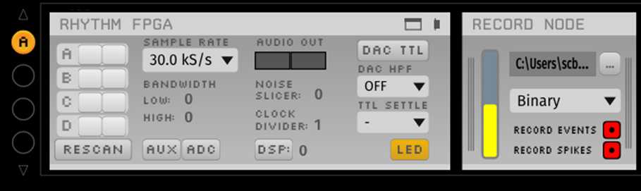
Open the Open Ephys GUI.
Open Ephys GUI must have
(1) an Acquisition Board node followed immediately by
(2) a Recording Node, saving the raw data in a binary format.
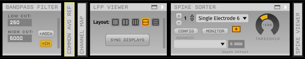
Optional: downstream in the processors graph, you can include a bandpass filter, common average reference node, LFP viewer, spike sorter, and spike viewer to view channels with spikes online during the recording.
- Begin recording (press “Play” then “Record”).
- After 15 minutes of baseline recording, begin the infusion (
1.5µLat0.1µL/min, taking 15 minutes). - After the infusion is complete, continue recording for
1h 30m 0s, then stop the recording. - Advance the electrodes by
26. - Return the mouse to its home cage to recover, and wait 2 weeks before recording again.
