Post-Surgical Dissection of Cervix
Stephen Fisher, Marielena Grijalva, Rong Guo, Sarah A Johnston, Hieu Nguyen, John Renz, Jean G Rosario, Steven Rudich, Brian Gregory, Junhyong Kim, Kate O'Neill
Abstract
This protocol describes dissection of the ectocervix and endocervix in preparation for 10X Visium, 10X Multiomics, pathology review, and biobanking. The cervix is the cylindrical lower part of the uterus which connects the uterus to the vagina. It is typically 3-4cm in length is largely fibromuscular in structure. The endocervical canal is lined with a thin layer of glandular epithelium. In this protocol we debulk the fibromuscular portion of the cervix for endocervical samples in order to ensure cells lining the endocervical canal are represented.
Before start
Dilute phosphate buffered saline (Gibco; 14200-075) to 1X with nuclease-free water.
Steps
Dissection of Ectocervix
Remove uterus from ice and dry the cervix.
Using a large grossing knife with a fresh blade, remove the most distal portion (1cm length) of the cervix which contains the ectocervix
Place the remainder of the cervix and uterus back on ice.
Weigh and measure this distal portion of cervix.
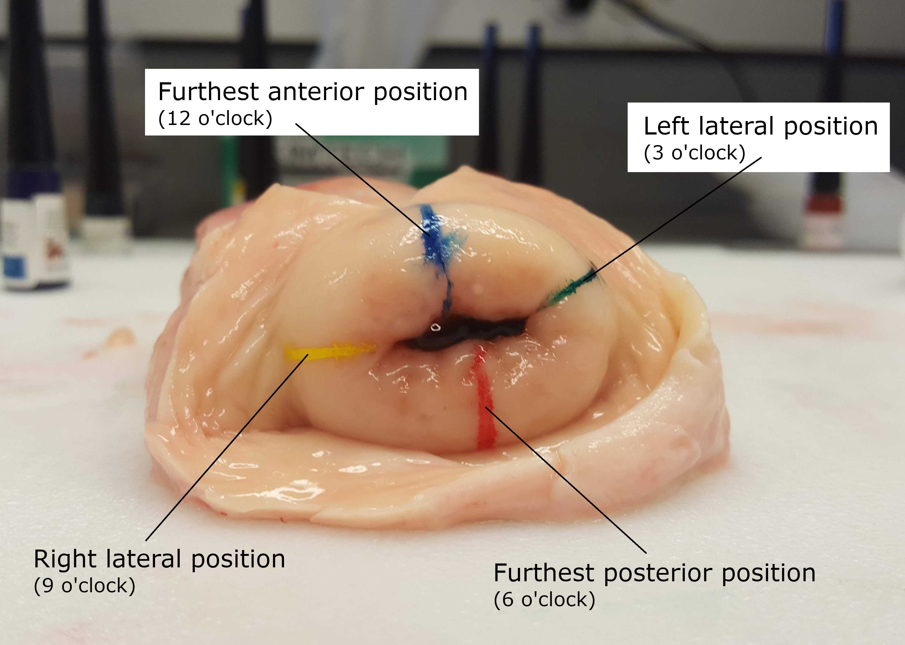
Lay down cervix with the external cervical os facing up, with the 12 o’clock anterior position furthest away from you.

Dry the ectocervix and label with marking dyes at 12 o’clock, 3 o’clock, 6 o’clock, and 9 o’clock.
Moving clockwise, slice the cervix vertically from 12 to 6 o’clock and horizontally from 9 to 3 o’clock so there are 4 equally sized pieces.
Divide each piece into four additional sections radially.
Using disposable weigh boats, weigh each tissue specimen.
For later 3D anatomical positioning, track the quadrant and radial section of each piece of tissue.
Tissue can be processed with protocols OCT-Embedded Tissue Preparation, Tissue Fixation Preparation, or Snap-Frozen Tissue Preparation depending on the desired downstream processing.
Dissection of Endocervix
Remove uterus and remaining cervix from ice and dry.
Using a large grossing knife with a fresh blade, remove the most distal 1cm of the cervix (previously the middle 1cm of cervix, prior to removal of ectocervical sample).
Return uterus to ice.
Trim ~1/2 of the eccentric cervical stroma.
Dry the endocervix and mark using marking dyes at 12 o’clock, 3 o’clock, 6 o’clock, and 9 o’clock.
Lay the cervix down with the external cervical os facing up, with the 12 o’clock anterior position furthest away from you.
Moving clockwise, slice the cervix in sections as was done above with ectocervix.
Divide each section into 4 additional sections radially.

Using disposable weigh boats, take the weight of each tissue specimen.
For later 3D anatomical positioning, track the quadrant and radial section of each piece of tissue.
Tissue can be processed with protocols OCT-Embedded Tissue Preparation, Tissue Fixation Preparation, or Snap-Frozen Tissue Preparation depending on the desired downstream processing.

