Pharmacological treatment with corticoid or hyaluronic acid injections into subtalar joint via lateral access
Fernanda Ferreira Gomes, Isnar M. de Castro Junior, João Antônio M. Guimarães, Aline Cordeiro
Abstract
Pharmacological treatment, such as corticoid or hyaluronic acid intra-articular injections, is consistently used for osteoarthritis cases in which conservative treatment failed. The use of intra-articular injections aims to decrease pain and restore function, at least partially and temporarily [1]. Nonetheless, the indication for foot joint injection is restricted, especially for the subtalar joint, since there is a lack of reports which provide technical information on this subject.
This protocol provides a step-by-step guide for performing subtalar intra-articular injection, which is a simple procedure that consists of inserting a needle in the subtalar joint to dispense pharmacological treatment directly in contact with the tissues committed by osteoarthritis. Since the intra-articular injection technique is invasive, there is a need to be cautious about performance to minimize risks of infection, discomfort, lesions and pain. For this, orthopedists might employ some methodological precautions as well as take special care regarding joint access.
The lateral route is easy to perform since it uses the anterior edge of the lateral malleolus tip as a landmark. Beyond that, there are no neurovascular bundles or tendons in the needle's path. However, in our literature searches, we did not identify records considering this route, except for previous articles by our group [2,3], which showed positive effects of corticoid and principally hyaluronic acid injections. Thus, we point the lateral access as a safe route, reinforcing the need to disclose this technique.
Before start
In a medical consultation previously to the procedure day, the patient might receive counseling regarding the benefits of intra-articular injections, information about the protocol scheme and the average duration of the medication effects. Beyond that, the patient should be warned about the risks of the treatment.
Steps
Patient preparation and positioning
Wash the patient's foot with degerming chlorhexidine and saline solution abundantly using a basin to store the liquid;
Dispose of the washing liquid in a suitable place;
Dry the patient's foot with a sterile compress;
Place the patient in lateral decubitus, with ipsilateral knee flexion between 80 and 90°;
Position the contralateral knee flexed at 45°;
Place the adipose cushion in the patient's ipsilateral medial malleolus region to promote subtalar inversion;
Wear sterile gloves;
Perform asepsis with sterile gauze and 70% alcohol or alcoholic chlorhexidine at the infiltration site;
Local anesthesia
Palpate the tip of the lateral malleolus;
Apply 1 ml of lidocaine to the skin and subcutaneous tissue slightly anterior to the lateral malleolus in various directions, according to the region marked in red (Figure 1A), after aspirating the syringe, making sure it is not inside a blood vessel;
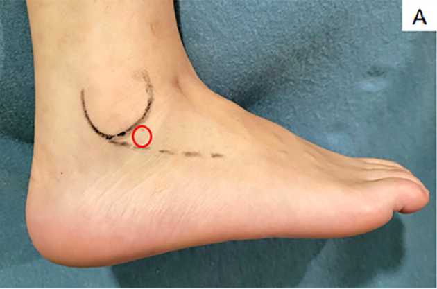
Make circular movements with a gauze, performing compression at the application site, to spread the anesthetic and stop the bleeding;
Medication preparation
Gently mix and aspirate the medication (hyaluronic acid and/or corticoid) directly from the manufacturer's vial(s);
For the subtalar joint, isolated drug administration usually composes a 2ml injection. However, the combined hyaluronic acid and corticoid maximum volume must not exceed 4 ml (Table 1 at Guidelines & Warnings ).
Subtalar intrarticular injection
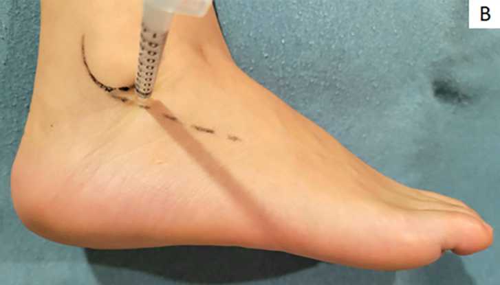
Palpate the tip of the lateral malleolus again to confirm the position where the intra-articular injection will be performed;
Introduce the needle attached to a 3 ml syringe in the region marked in the site pointed in red or use the needled syringe supplied by the manufacturer containing hyaluronic acid or its association with corticoid, directing the needle towards the posterior facet (Figure 1B). The needle angle of attack is 90°, but it may be necessary to change angle to find the joint cleft, considering that some patients have significant narrowing or deformity of the bone contour. Assessing radiography before the procedure is very important. The content must enter without resistance and discomfort for the patient. If this does not happen, remove the needle, reposition it, and reinsert it again following the same procedure;
After applying all the contents, remove the needle and compress the site with gauze for about two minutes to stop the bleeding and reduce the formation of a hematoma;
Perform inversion and eversion movements in the subtalar to spread the medicine.
