Nucleus-highlighting (terminal/lethal) staining of adherent live U2-OS cells by Erythrosine B

Misha Koksharov

Published: 2022-01-20 DOI: 10.17504/protocols.io.bvfjn3kn

Abstract

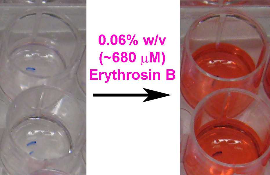
Erythrosine B (tetraiodofluorescein; ErB) is a negatively charged viability stain commonly used to assess cell viability in a hemocytometer or in adherent mammalian cell cultures. Being considered membrane-impermeable, it preferentially stains dead cells that have lost their membrane integrity (pink color within 1 min of treatment). It is often recommended over Trypan Blue staining for providing a faster and more reliable dead cell staining and for being a generally non-toxic reagent. While exposure to Trypan Blue is toxic to mammalian cells, ErB is often stated to be relatively harmless and suitable for non-terminal cell staining. However, Erythrosine B actually enters live cells and eventually stains them lethally but the speed of this process varies considerably between cell lines and can take from several minutes (U2-OS) to more than 30 minutes (HEK293T).

In the presence of 0.06% w/v (or higher) ErB in serum-free media the adherent U2-OS cells get efficiently stained after 10 min resulting in a pronounced nuclear and a faint cytoplasmic staining while retaining the general cell morphology and adherence. This can be used for a fast terminal staining and counting of adherent U2-OS cells or other cell lines that interact with ErB in the same way.

Lower ErB concentrations in serum-free media often lead to a fast cell disintegration and loss of nuclei in U2-OS cells without staining. The discussed possible destructive and toxic effects of Erythrosine B on mammalian cells should be taken into account when using it as a vital dye. The presence of serum in the media temporarily protects live U2-OS cells from the toxic action of ErB (at concentrations below 0.1% w/v), so it can be recommended when staining dead cells to avoid affecting the live ones.

Before start

Prepare the required stock solution and media. Read the Guidelines & Warnings section describing experimental examples and the literature background of the method.

Steps

Cell staining protocol steps

1.

Grow U2-OS cells in a desired vessel.

Adherent U2-OS cells grown on a surface of 24-well plate (TC-treated transparent polystyrene plate).

Note
U2-OS mammalian cell line demonstrates a strong adherence to the growth substrate and can grow well in a variety of media of different nutritional composition (DMEM, McCoy's 5A, F10, F12, MCDB105, etc) in the presence of 1-10 % serum.Most commonly it is grown in DMEM or McCoy's 5A medium in the presence of 5-10% FBS.

2.

Remove the culture media from the vessel (cell culture plate or well) with adherent U2-OS cells.

Note
It is also possible to use the starting serum-containing growth media without a change but the final concentration of Erythrosin B will need to be increased to 0.2% w/v or more to achieve the same results. Otherwise, serum proteins bind ErB blocking or reducing most of the described effects on live cells.

3.

Gently add a serum-free cell culture media or buffer (preferably maintaining pH 7.2-7.6 in the absence of the CO2 atmosphere) to cover the cells.

Note
In principle, any buffer or media not affecting live cells in a short time scale is suitable, since the staining is terminal and short. A physiological buffer with calcium and magnesium or a cell culture medium similar to the one used for cell growth can be a good choice.It is convenient to have a transparent media without phenol redIf using a cell culture medium, it is useful if it's buffered to have pH 7-7.6 in the absence of CO2 atmosphere (like MCDB media or regular cell culture media formulations without sodium bicarbonate) if cells are to be handled outside cell culture incubator. However, the described terminal staining works similar regardless of this point.

4.

Add a small volume of 6% w/v (~6.8 mM) Erythrosin B stock (in water) to the media above cells to achieve the final 0.06% concentration (1:100 dilution). Mix by gently shaking the plate.

Note
In serum-containing media the concentration of ErB should be increased to 0.2% (with 5% FBS) or more to achieve the same results.0.006% ErB is not sufficient to stain live U2-OS cells but leads to cell damage (see the Guidelines section); this concentration still stains dead cells.

5.

Incubate the plate for 10-15 min in a cell culture incubator (37°C).

Note
During this time, living U2OS cells will uptake ErB from the media leading to a faint pink coloration of the cytosol with a pronounced pink staining of the nucleus.

6.

The medium can be exchanged to a fresh transparent one or removed.

Cells can now be imaged in phase contrast or brightfield: nuclei become intensely pink while cytosol remains quite faint. If placed under ErB-free medium, cells will loose some of the accumulated ErB.

Image of (initially) live U2OS cells stained with ErB. Left: phase contrast image; Right: brightfield image. Cells were grown and stained in wells of a 24-well TC-treated polystyrene plate.

Post-protocol notes

7.

Note
A flat shape and large size of U2OS cells generally results in a good visual separation of pink nuclei between neighboring cells.Stained cells loose their viability but remain fixed.The use of ErB concentrations below 0.01% leads to the separation of nuclei from cells and disintegration of many U2-OS cells rather than staining and fixation.

Note
REFERENCES 1. ATCC (2020) ATCC® Animal cell culture guide. 1. ATCC (2020) ATCC® Animal cell culture guide. https://www.atcc.org/-/media/resources/culture-guides/animal-cell-culture-guide.pdf2. Avelar-Freitas, B. A., Almeida, V. G., Pinto, M. C., Mourão, F. A., Massensini, A. R., Martins-Filho, O. A., Rocha-Vieira, E., & Brito-Melo, G. E. (2014). Trypan blue exclusion assay by flow cytometry. Brazilian journal of medical and biological research , 47(4), 307–315. 2. Avelar-Freitas, B. A., Almeida, V. G., Pinto, M. C., Mourão, F. A., Massensini, A. R., Martins-Filho, O. A., Rocha-Vieira, E., & Brito-Melo, G. E. (2014). Trypan blue exclusion assay by flow cytometry. Brazilian journal of medical and biological research, 47(4), 307–315. https://doi.org/10.1590/1414-431X201434373. Chan, L. L., Rice, W. L., & Qiu, J. (2020). Observation and quantification of the morphological effect of trypan blue rupturing dead or dying cells. PloS one , 15 (1), e0227950. 3. Chan, L. L., Rice, W. L., & Qiu, J. (2020). Observation and quantification of the morphological effect of trypan blue rupturing dead or dying cells. PloS one, 15(1), e0227950. https://doi.org/10.1371/journal.pone.02279504. Feenstra, R. P., & Tseng, S. C. (1992). What is actually stained by rose bengal?. Archives of ophthalmology , 110(7), 984–993. 4. Feenstra, R. P., & Tseng, S. C. (1992). What is actually stained by rose bengal?. Archives of ophthalmology, 110(7), 984–993. https://doi.org/10.1001/archopht.1992.010801900900355. Franke, J. D., Braverman, A. L., Cunningham, A. M., Eberhard, E. E., & Perry, G. A. (2020). Erythrosin B: a versatile colorimetric and fluorescent vital dye for bacteria. BioTechniques , 68 (1), 7–13. 5. Franke, J. D., Braverman, A. L., Cunningham, A. M., Eberhard, E. E., & Perry, G. A. (2020). Erythrosin B: a versatile colorimetric and fluorescent vital dye for bacteria. BioTechniques, 68(1), 7–13. https://doi.org/10.2144/btn-2019-00666. Ganesan, L., Margolles-Clark, E., Song, Y., & Buchwald, P. (2011). The food colorant erythrosine is a promiscuous protein-protein interaction inhibitor. Biochemical pharmacology , 81(6), 810–818. 6. Ganesan, L., Margolles-Clark, E., Song, Y., & Buchwald, P. (2011). The food colorant erythrosine is a promiscuous protein-protein interaction inhibitor. Biochemical pharmacology, 81(6), 810–818. https://doi.org/10.1016/j.bcp.2010.12.0207. Ganesan, L., & Buchwald, P. (2013). The promiscuous protein binding ability of erythrosine B studied by metachromasy (metachromasia). Journal of molecular recognition , 26(4), 181–189. 7. Ganesan, L., & Buchwald, P. (2013). The promiscuous protein binding ability of erythrosine B studied by metachromasy (metachromasia). Journal of molecular recognition, 26(4), 181–189. https://doi.org/10.1002/jmr.22638. Giulian, D., & Diacumakos, E. G. (1977). The electrophysiological mapping of compartments within a mammalian cell. The Journal of cell biology , 72 (1), 86–103. 8. Giulian, D., & Diacumakos, E. G. (1977). The electrophysiological mapping of compartments within a mammalian cell. The Journal of cell biology, 72(1), 86–103. https://doi.org/10.1083/jcb.72.1.869. Ham, R. G., & McKeehan, W. L. (1979). Media and growth requirements. Methods in enzymology , 58 , 44–93. 9. Ham, R. G., & McKeehan, W. L. (1979). Media and growth requirements. Methods in enzymology, 58, 44–93. https://doi.org/10.1016/s0076-6879(79)58126-910. Kim, S. I., Kim, H. J., Lee, H. J., Lee, K., Hong, D., Lim, H., Cho, K., Jung, N., & Yi, Y. W. (2016). Application of a non-hazardous vital dye for cell counting with automated cell counters. Analytical biochemistry , 492 , 8–12. 10. Kim, S. I., Kim, H. J., Lee, H. J., Lee, K., Hong, D., Lim, H., Cho, K., Jung, N., & Yi, Y. W. (2016). Application of a non-hazardous vital dye for cell counting with automated cell counters. Analytical biochemistry, 492, 8–12. https://doi.org/10.1016/j.ab.2015.09.01011. Krause, A. W., Carley, W. W., & Webb, W. W. (1984). Fluorescent erythrosin B is preferable to trypan blue as a vital exclusion dye for mammalian cells in monolayer culture. The journal of histochemistry and cytochemistry , 32 (10), 1084–1090. 11. Krause, A. W., Carley, W. W., & Webb, W. W. (1984). Fluorescent erythrosin B is preferable to trypan blue as a vital exclusion dye for mammalian cells in monolayer culture. The journal of histochemistry and cytochemistry, 32(10), 1084–1090. https://doi.org/10.1177/32.10.609053312. Lee, Y. C., Park, C. K., Kim, M. S., & Kim, J. H. (1996). In vitro study for staining and toxicity of rose bengal on cultured bovine corneal endothelial cells. Cornea , 15 (4), 376–385. 12. Lee, Y. C., Park, C. K., Kim, M. S., & Kim, J. H. (1996). In vitro study for staining and toxicity of rose bengal on cultured bovine corneal endothelial cells. Cornea, 15(4), 376–385. https://doi.org/10.1097/00003226-199607000-0000813. LOEWENSTEIN, W. R., & KANNO, Y. (1963). SOME ELECTRICAL PROPERTIES OF A NUCLEAR MEMBRANE EXAMINED WITH A MICROELECTRODE. The Journal of general physiology , 46 (6), 1123–1140. 13. LOEWENSTEIN, W. R., & KANNO, Y. (1963). SOME ELECTRICAL PROPERTIES OF A NUCLEAR MEMBRANE EXAMINED WITH A MICROELECTRODE. The Journal of general physiology, 46(6), 1123–1140. https://doi.org/10.1085/jgp.46.6.112314. Mathavan, V. M., Boh, B. K., & Tayyab, S. (2009). Characterization of erythrosine B binding to bovine serum albumin and bilirubin displacement. Indian journal of biochemistry & biophysics , 46(4), 325–331. 14. Mathavan, V. M., Boh, B. K., & Tayyab, S. (2009). Characterization of erythrosine B binding to bovine serum albumin and bilirubin displacement. Indian journal of biochemistry & biophysics, 46(4), 325–331. https://pubmed.ncbi.nlm.nih.gov/19788065/15. Matzke And, M. A., & Matzke, A. J. M. (1985). Potential difference across the nuclear membrane: a regulator of gene expression?. Journal of Bioelectricity , 4 (2), 461-480. 15. Matzke And, M. A., & Matzke, A. J. M. (1985). Potential difference across the nuclear membrane: a regulator of gene expression?. Journal of Bioelectricity, 4(2), 461-480. https://doi.org/10.3109/1536837850903326616. Milo R. (2013). What is the total number of protein molecules per cell volume? A call to rethink some published values. BioEssays , 35 (12), 1050–1055. 16. Milo R. (2013). What is the total number of protein molecules per cell volume? A call to rethink some published values. BioEssays, 35(12), 1050–1055. https://doi.org/10.1002/bies.20130006617. Nguyen, P., Clark, E. R., Ananthakrishnan, S., Lenz, K., & Canavan, H. E. (2020). How to select the appropriate method(s) of cytotoxicity analysis of mammalian cells at biointerfaces: A tutorial. Biointerphases , 15 (3), 031201. 17. Nguyen, P., Clark, E. R., Ananthakrishnan, S., Lenz, K., & Canavan, H. E. (2020). How to select the appropriate method(s) of cytotoxicity analysis of mammalian cells at biointerfaces: A tutorial. Biointerphases, 15(3), 031201. https://doi.org/10.1116/6.000013618. Sablin, N. V., Gerasimova, M. A., & Nemtseva, E. V. (2016). Spectral changes of erythrosin B luminescence upon binding to bovine serum albumin. Russian Physics Journal , 58 (12), 1797-1803. 18. Sablin, N. V., Gerasimova, M. A., & Nemtseva, E. V. (2016). Spectral changes of erythrosin B luminescence upon binding to bovine serum albumin. Russian Physics Journal, 58(12), 1797-1803. https://doi.org/10.1007/s11182-016-0719-619. Scott, M. F., & Merrett, H. J. (1995). Evaluation of Erythrocin B as a Substitute for Trypan Blue. In Animal Cell Technology: Developments Towards the 21st Century (pp. 1133-1139). Springer, Dordrecht. 19. Scott, M. F., & Merrett, H. J. (1995). Evaluation of Erythrocin B as a Substitute for Trypan Blue. In Animal Cell Technology: Developments Towards the 21st Century (pp. 1133-1139). Springer, Dordrecht. https://doi.org/10.1007/978-94-011-0437-1_17820. Soedjak H. S. (1994). Colorimetric micromethod for protein determination with erythrosin B. Analytical biochemistry , 220 (1), 142–148. 20. Soedjak H. S. (1994). Colorimetric micromethod for protein determination with erythrosin B. Analytical biochemistry, 220(1), 142–148. https://doi.org/10.1006/abio.1994.131021. Tsaousis, K. T., Kopsachilis, N., Tsinopoulos, I. T., Dimitrakos, S. A., Kruse, F. E., & Welge-Luessen, U. (2013). Time-dependent morphological alterations and viability of cultured human trabecular cells after exposure to Trypan blue. Clinical & experimental ophthalmology , 41 (5), 484–490. 21. Tsaousis, K. T., Kopsachilis, N., Tsinopoulos, I. T., Dimitrakos, S. A., Kruse, F. E., & Welge-Luessen, U. (2013). Time-dependent morphological alterations and viability of cultured human trabecular cells after exposure to Trypan blue. Clinical & experimental ophthalmology, 41(5), 484–490. https://doi.org/10.1111/ceo.12018

10.

推荐阅读

Nature Protocols
Protocols IO
Current Protocols
扫码咨询