Non-Photochemical Quenching (NPQ) Relaxation Analysis by CF imager FluorCam FC800-331 and FluorCam 7 Software
Lynn Doran, Steven J Burgess
Abstract
For assessment of Non-Photochemical Quenching (NPQ) relaxation in leaf discs using chlorophyll fluorescence on FluorCam FC800-331 and FluorCam 7 Software under 10 minutes of low light (50 umolm-2s-1) to activate rubisco, 15 minutes of high light (1600 umolm-2s-1) to activate NPQ without causing photodamage, and 45 minutes of NPQ relaxation under low light (50 umolm-2s-1). Fluorescence measurements using saturating pulses are taken every 2.5 minutes during high light and for the first two time points after return to low light and every 5 minutes thereafter.
Before start
Prepare samples as described in "Sampling leaf tissue for analysis of NPQ Relaxation using Technologica Chlorophyll Fluorescence Imager Data. V.2".
If it is the start of an experiment set, different plant or tissue type, modifications have been made to the instrument, or modifications have been made to the protocol, prepare an additional sample plate to be dark adapted. This additional plate will be used to set the initial camera settings. Typically, once the hardware and software settings are adjusted, similar samples can be analyzed across different experiments or days using the same settings and additional plates will not need to be prepared each day. For focusing and zooming the camera, the size calibration standard can be used.
Read Photon Systems Instruments Closed Fluorcam Manual for detailed instrument setup and operation instructions. For quick start instructions, reference the PSI Quick Start Guide.
Reserve instrument using the Google Calendar. Access to the equipment reservation calendars can be requested through the IGB GEGC lab manager.
Steps
Setting up the CF Imager
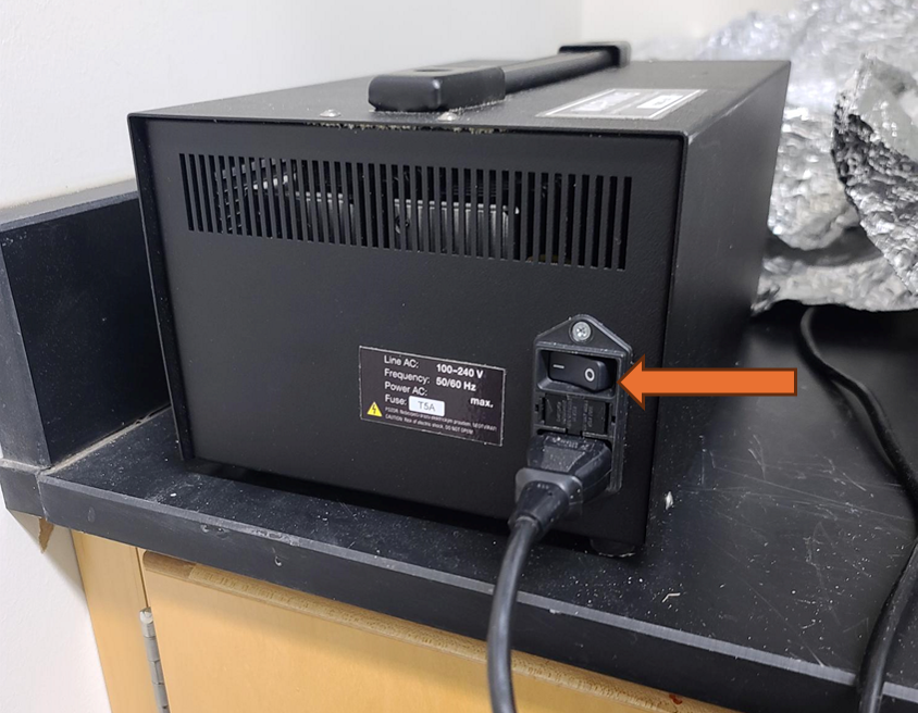
Turn on the black power supply box. The switch is on when the - symbol is depressed and the O symbol is lifted.
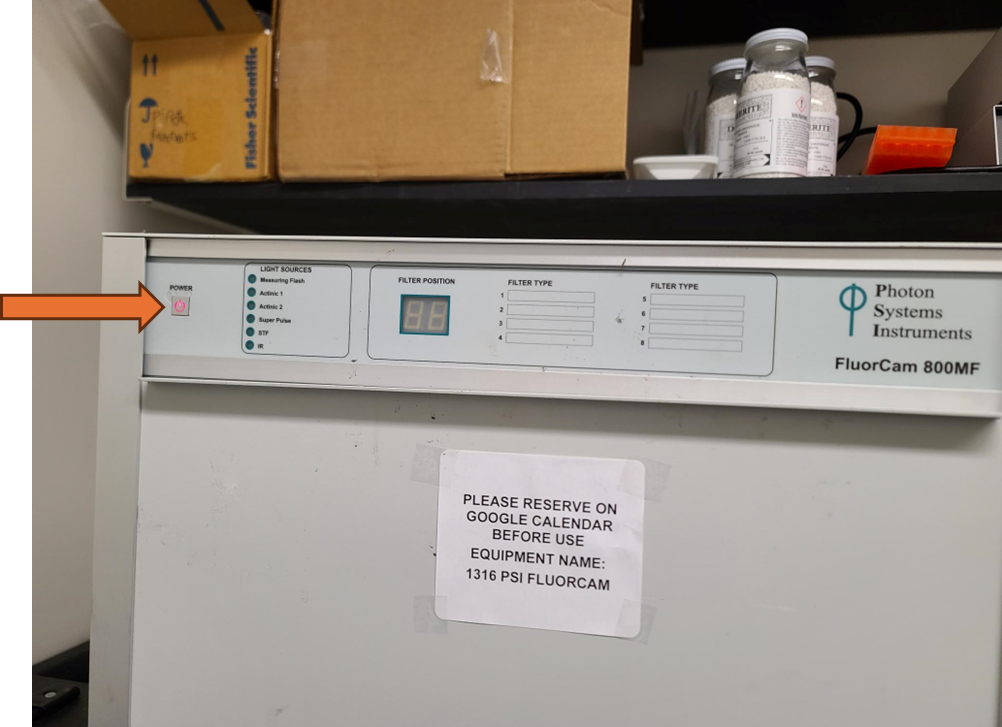
Turn on the Fluorcam CF Imager using the power button in the top left. The button will illuminate red when it is on.
Open the software "Fluorcam7" by double clicking on the app or on the leaf icon.
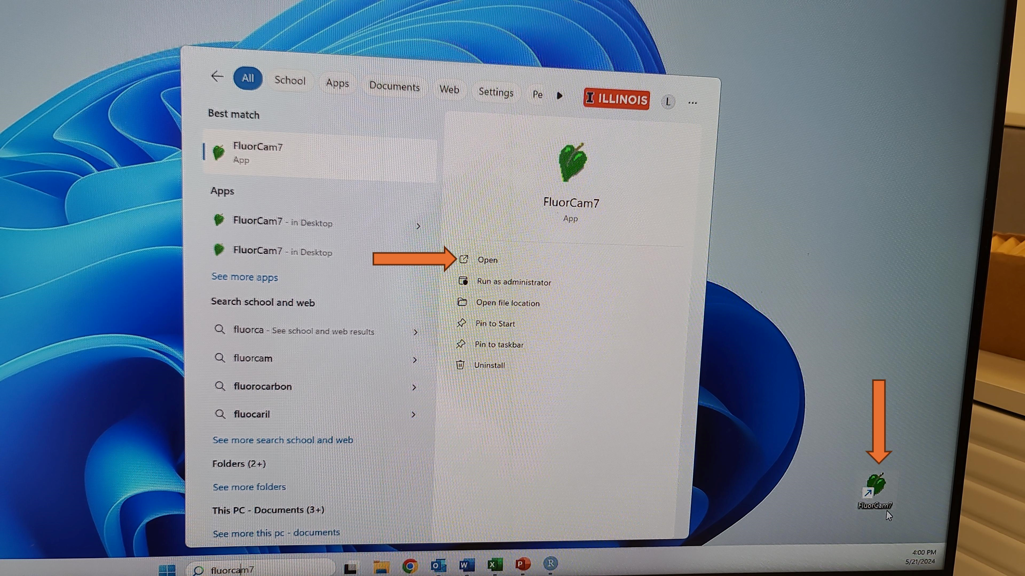
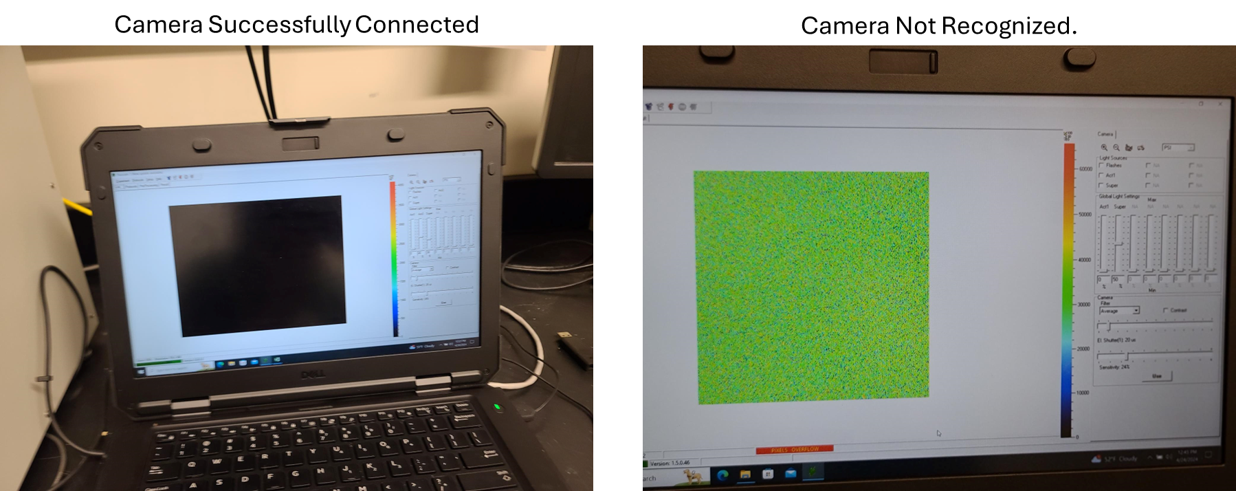
The black power supply and the white Fluorcam Instrument must both be on and fully booted for the software to recognize the Fluorcam cameras. If a black home screen appears when the software is open, the instrument is properly connected. If a yellow static screen appears, close the software, ensure both power buttons are on, wait a few minutes, and reopen the software.
If camera has already been focused, desired plate masks have been made, and the camera shutter and sensitivity settings have previously been prepared for this experiment, hardware configuration, and plant and tissue type, skip sections "Camera Focus- Performed Once at Start of Experiment", "Optional: Size Calibration (Necessary for Object Size Reporting and Using Predefined Plate Maps)", and Adjusting Camera Shutter and Sensitivity Settings - Performed Once at Start of Experiment and go straight to section "Analysis".
Camera Focus- Performed Once at Start of Experiment
Place the size calibration standard sheet in the middle of the shelf for focus adjustments.
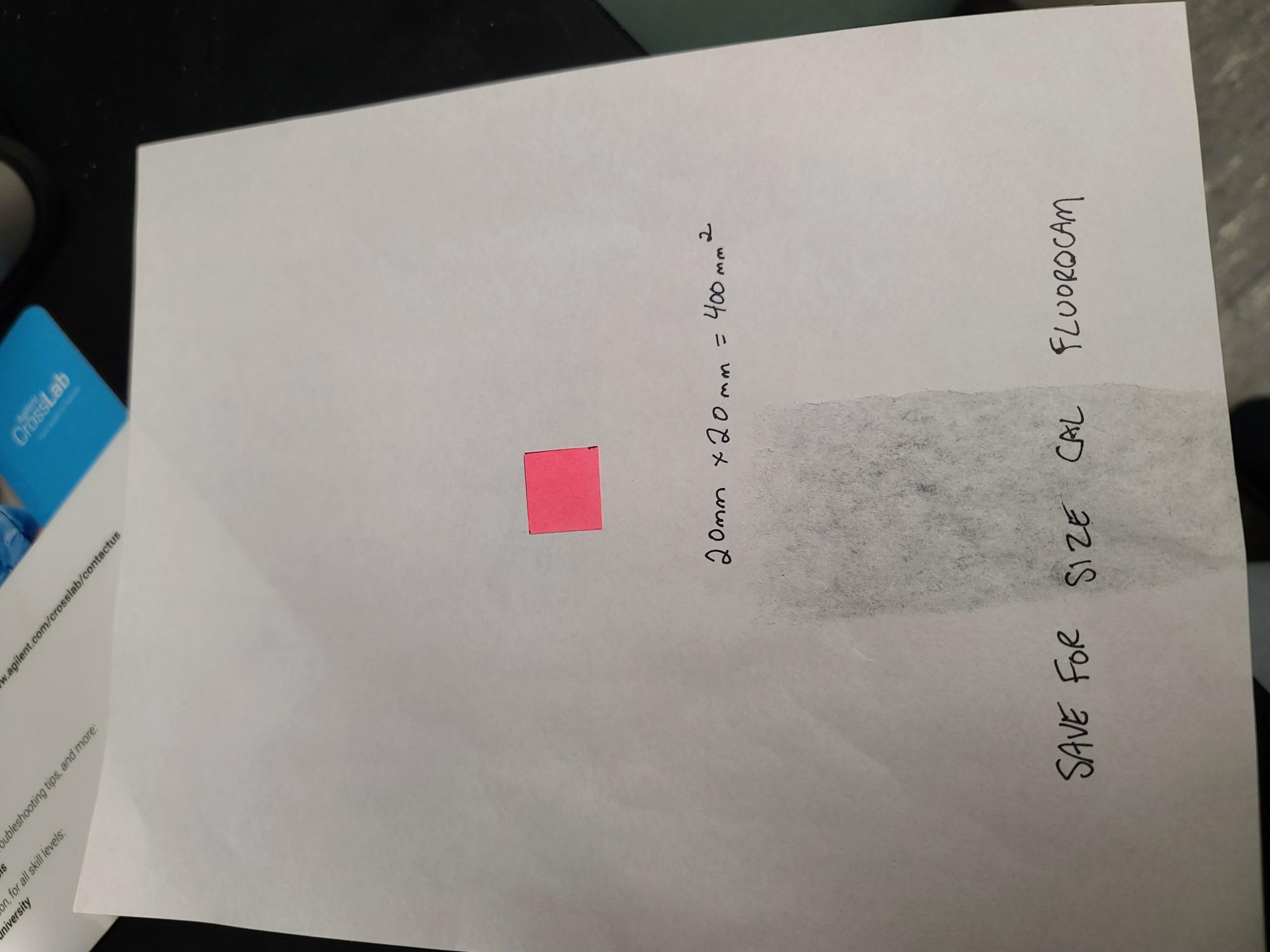
If the size calibration standard that comes with the instrument is not available, a pink fluorescent post it note can be measured and cut to exactly 20 mm by 20 mm and taped to a piece of paper. Add text to the paper to provide a focal point for focusing the camera.
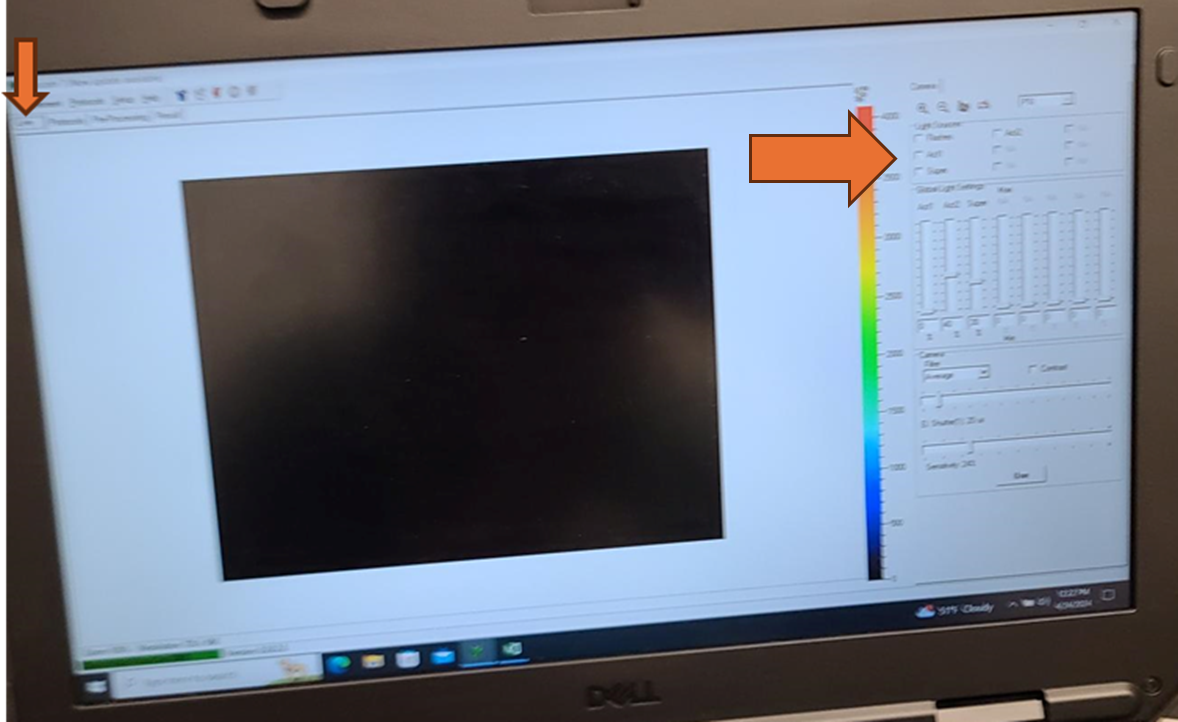
Turn on the white actinic light by selecting the checkbox next to "Act 2". Adjust the intensity of the Act 2 light source by using the slider bar or changing the percent intensity in the box below the slider bar until the text on the size calibration standard is visible.

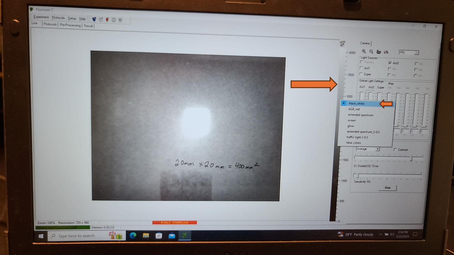
Change the color scale to black and white by right clicking on the color bar to the right of the live field of view and selecting "black_white".
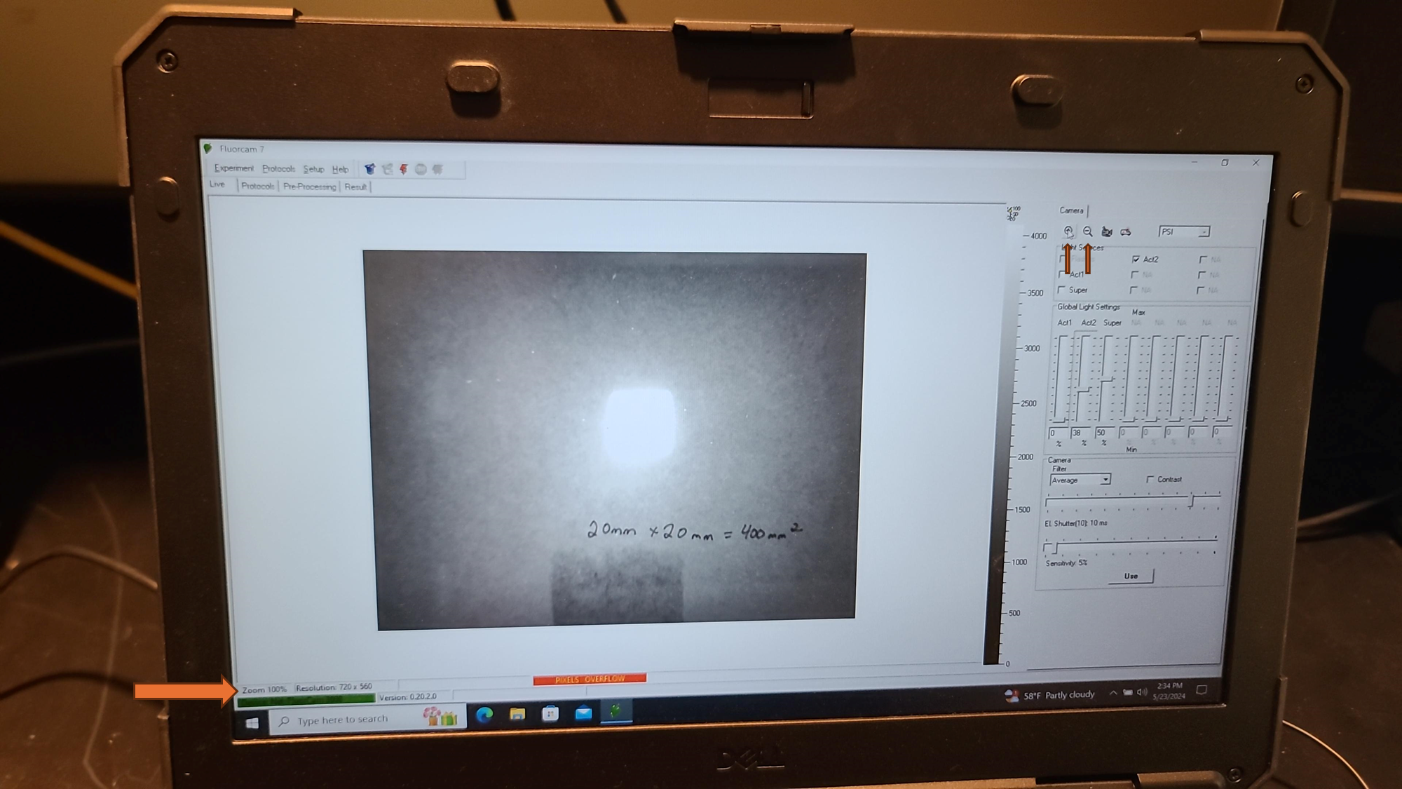
Set the zoom to 100% using the + and - signs in magnifying glasses at the top left of the control panel. The percent zoom is reported in the bottom left corner under the live field of view.
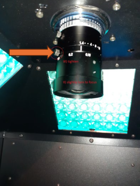
The focus needs to be manually adjusted on the camera inside the Fluorcam. The camera is located inside the Fluorcam box in the center of the LED light panels on the top of the box. Loosen the bottom of the two set screws by turning the screw counter clockwise to allow adjustment of the focus.
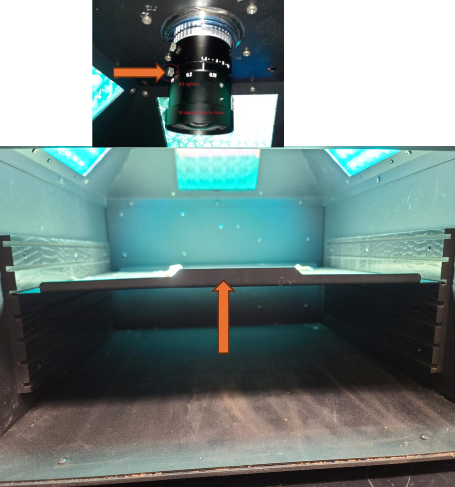
Watching the live field of view screen, slowly and gently twist the bottom portion of the camera until the lettering on the size calibration standard is clear.
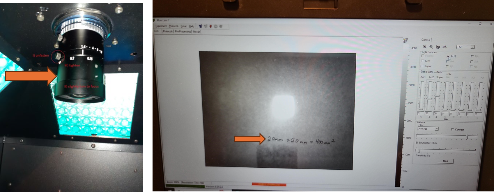
Fix the focus in place by tightening the bottom of the two set screws by turning clockwise until finger tight.
Once the focus is set, do not adjust it again for the remainder of the experiment. The level of absolute fluorescence signal could change when the focus of the camera is changed.
Optional: Size Calibration (Necessary for Object Size Reporting and Using Predefined Plate Maps)
If the size in millimeters of the objects measured is desired, then the size calibration must be done before each analysis. If the size calibration is being used to create a pre-processing mask (for example for a 96 well plate), it must be done before the analysis when the mask is created. Once the pre-processing mask is created, as long as the objects measured are the same size and distance from the camera, a size calibration is not needed before every analysis to use the mask.
Ensure all lights are manually turned off by unchecking boxes near Flashes, Act1, Super, and Act 2 in the upper right corner.
From the top toolbar, select "Setup". From the drop down menu, double click on "Size Calibration".
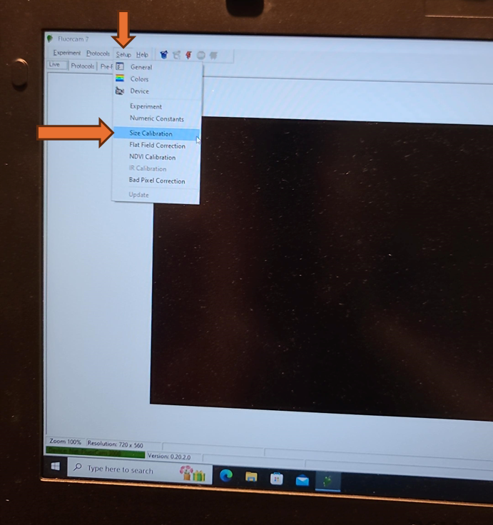
A command box with instructions to insert calibration plate will appear. Insert the size calibration standard and center the fluorescent square in the field of view. Then click "OK" in the command box.

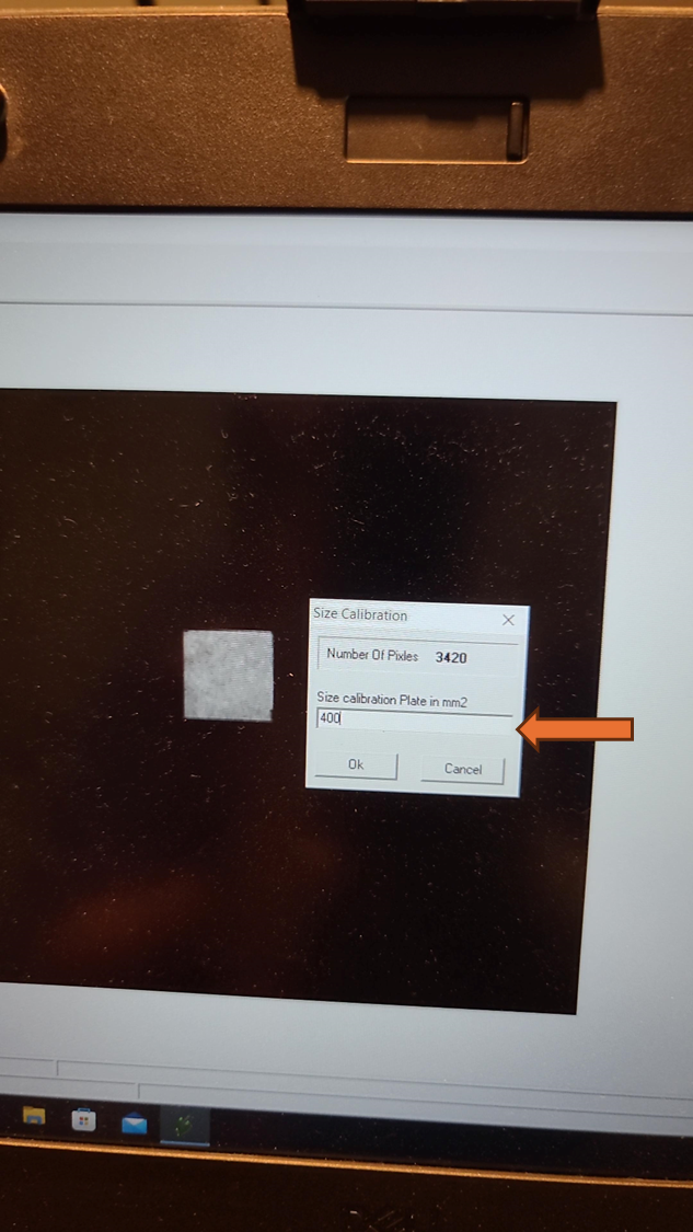
A second command box will appear showing the number of pixels measured. Enter the size of the Size Calibration Standard. This is the area of the shape measured. If a 20 mm by 20 mm size calibration standard was used, enter 400 into the mm2 box.
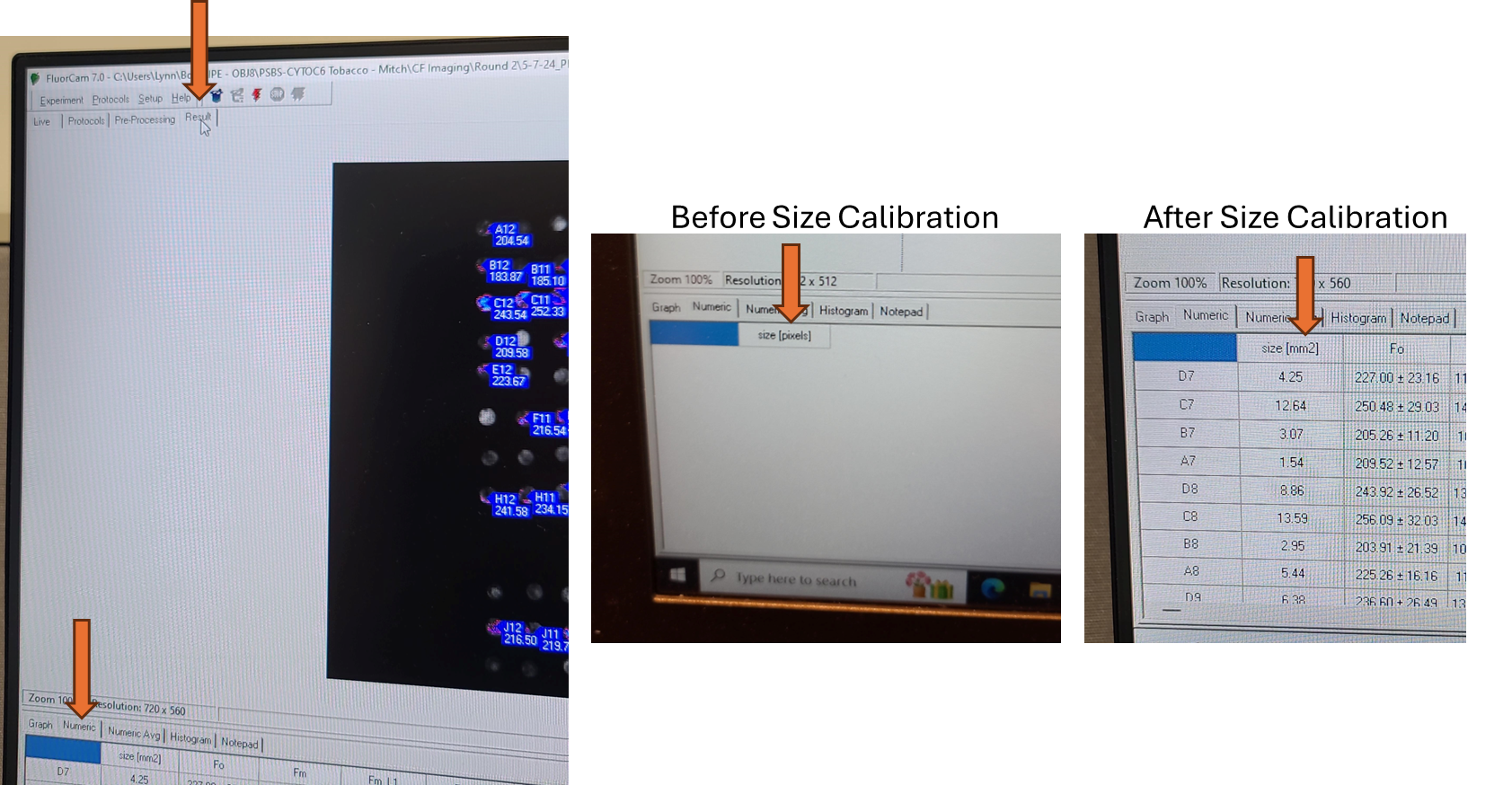
After completion of your analysis, the "Results" tab, "Numeric" results will now show sizes of identified objects in mm2 instead of pixels.
Adjusting Camera Shutter and Sensitivity Settings - Performed Once at Start of Experiment
The shutter and sensitivity settings need to be optimized once at the start of the experiment and then should not be changed again for consistent fluorescent readings across all sample analysis runs. The shutter and sensitivity settings may need to be adjusted if plant species, tissue type, distance from camera, plant health, or plant growth conditions change affecting the baseline emission of chlorophyll fluorescence.
The goal is to adjust the shutter and sensitivity settings high enough that the camera reliably measures fluorescence at all desired timepoints under all desired conditions while avoiding pixel overload that could cause inaccurate readings and minimize the amount of additional non-treatment actinic light exposure.
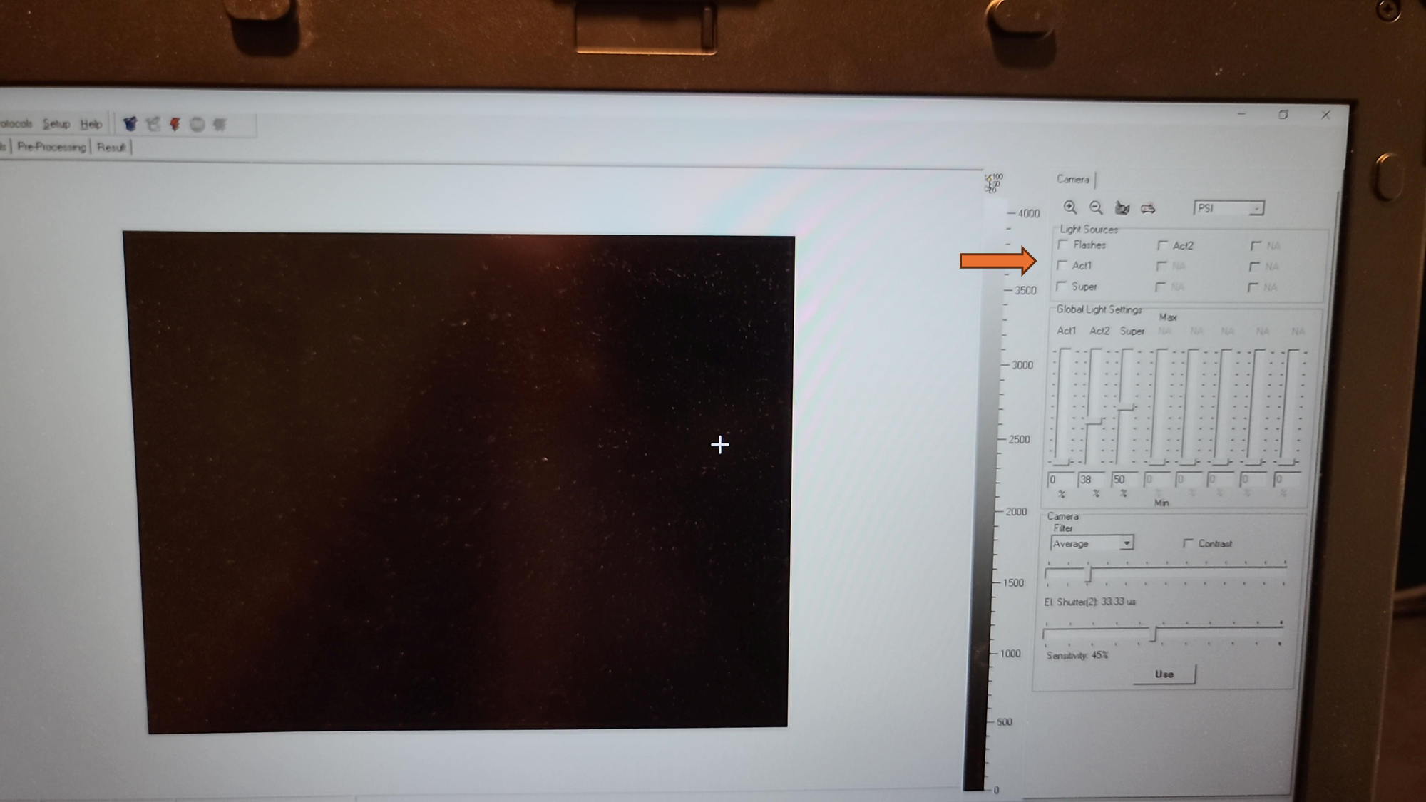
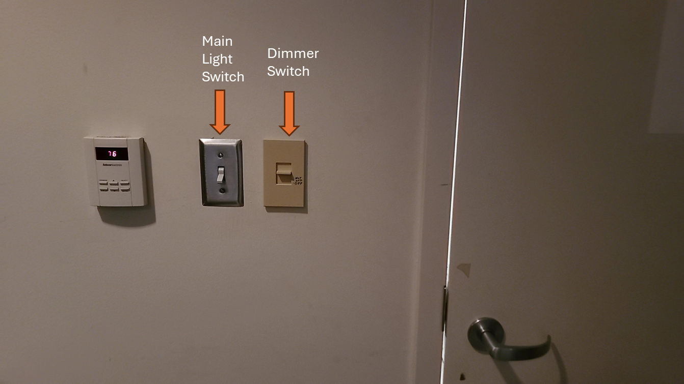
Turn off the primary overhead light and use the dimmer switch on the wall to set the lighting in the room as low as you can safely work in, with dark being ideal. Ambient light can affect shutter settings and the camera sensitivity. Leaf discs for analysis should have as limited light exposure prior to initial fluorescence measurement as possible.
Ensure all lights on the Fluorcam are manually turned off by unchecking boxes near Flashes, Act1, Super, and Act 2 in the upper right corner.
Remove any aluminum foil and parafilm from the additional sample plate that was prepared and dark adapted (from "Before Start" section), flip the plate upside down so the adaxial side of the leaf faces towards the Fluorcam camera, and place on the shelf with the corner of the plate snugly against the guardrails for the field of view.
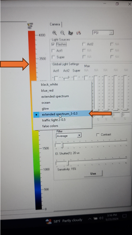
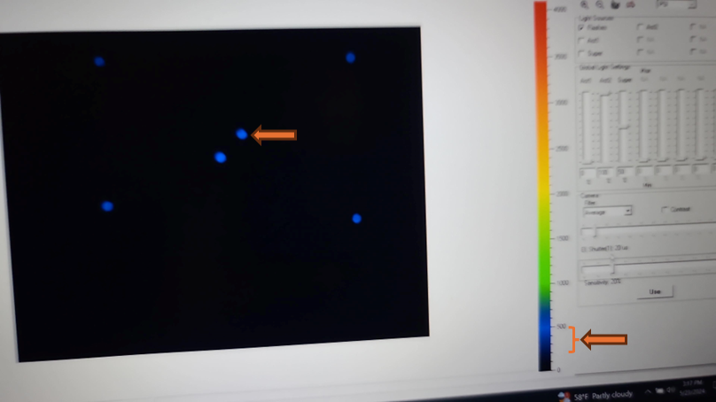
Change the color scale to "extended spectrum 3" by right clicking on the color bar to the right of the live field of view and selecting "extended spectrum_3-0.3".
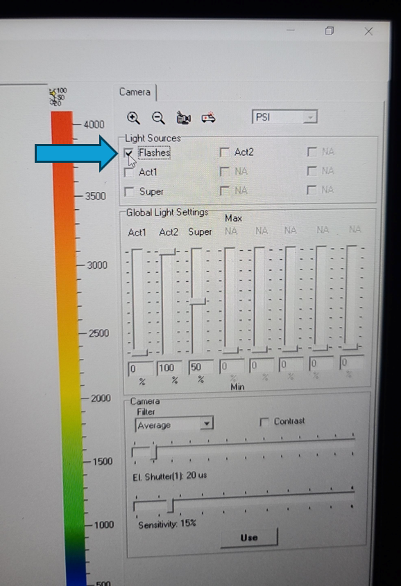
Turn on flashes by selecting check box next to "flashes" in upper right control window.
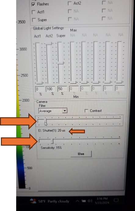
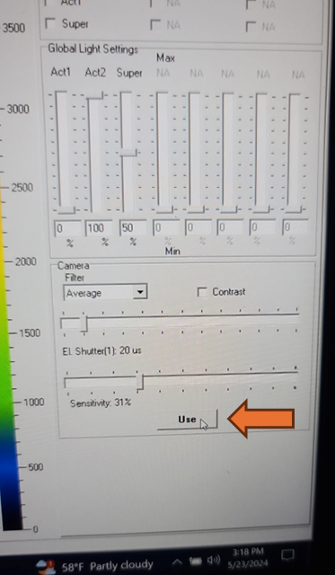
Set the EI shutter slider bar to 1 and adjust sensitivity slider bar until the leaf discs (or other plant tissue) is visible in the extended color scale and are a blue corresponding to between 200 and 500 on the color scale to the right of the live window. If it is not possible to get the tissue to appear on the highest sensitivity and EI shutter 1, increase EI shutter to 2 and repeat. The goal is to keep the EI shutter as low as possible to reduce unintended actinic light treatment.
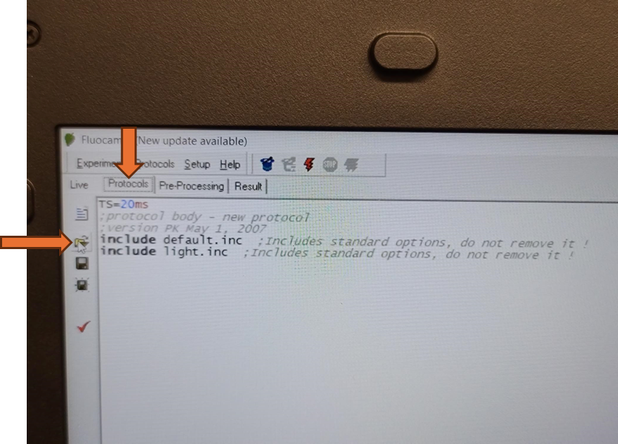
The updated EI shutter and sensitivity settings need to be imported into an existing protocol. Navigate to the protocols tab above the live field of view and use the open folder icon to select the desired protocol from the computer file system.
Protocol for NPQ relaxation:
VPZ-NPQ_50umol-10m_1600umol-15min_50umol-45min_annotated_v2.p
Alternatively, a pre-defined Fluorcam protocol can be selected and configured from the "Protocols" tab by selecting the wizard hat icon.
Once a protocol is successfully loaded, the coding section of the screen should populate. Be aware that the save and save as icons on the left side of this screen will only save modifications made to the loaded protocol. It will not save any of the size calibrations or camera settings in the live window.

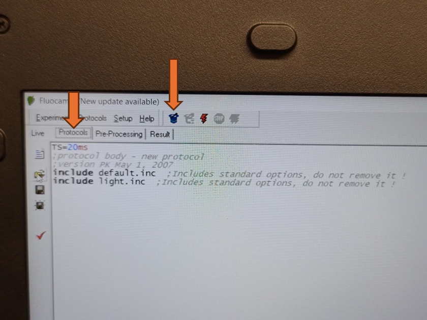
Once the desired protocol is loaded, navigate back to the live window by selecting the "Live" tab.
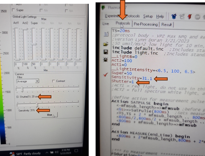
Update the EI shutter and sensitivity settings in the protocol by selecting the "Use" button on the right navigation pane.
Verify that the EI shutter and sensitivity settings were successfully imported into your protocol. Take note of the EI shutter and sensitivity numeric values on the Live window. Navigate to the protocols page using the "Protocol" tab at the top of the live field of view. Verify that the protocol code at the top of the method for Sensitivity and Shutter match the values that were selected on the Live window.
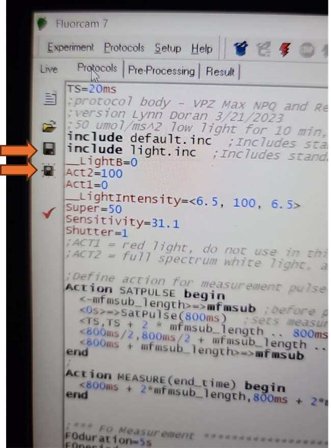
Save the updated protocol by selecting the save or save as icon on the Protocol tab. This will only save the updated protocol with EI shutter and sensitivity settings. The size calibration will not be saved until an analysis is run.
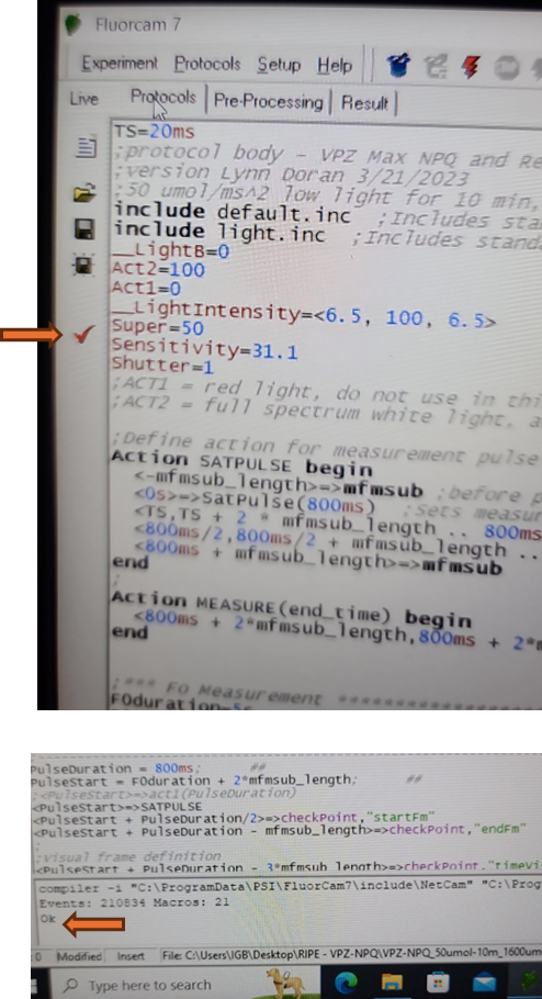
Verify that the protocol is correctly coded by selecting the check mark icon on the left side of the protocol tab. If the protocol is functional, the message box at the bottom of the protocol tab will output "OK". The instrument is now ready for experimental analysis.
Analysis
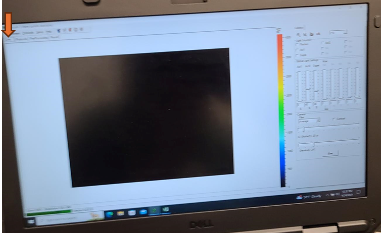
Navigate back to the live window by selecting the "Live" tab, ensure all instrument lights are off by unchecking flashes, Act1, Act2, and Super.
If applicable, remove the sample plate that was used for adjusting camera settings. It is no longer dark adapted and should not be used for real measurements.
Turn off the primary overhead light and using the dimmer switch on the wall, set the lighting in the room as low as you can safely work in, with dark being ideal. Ambient light can affect shutter settings and the camera sensitivity. Leaf discs for analysis should have as limited light exposure prior to initial fluorescence measurement as possible.
If used for dark adaptation, remove aluminum foil from sample plate.
If used, remove parafilm from plate edges. Humidity should be maintained for analysis by the sponges and any parafilm covering a leaf disc could interfere with light treatments and saturating pulses.
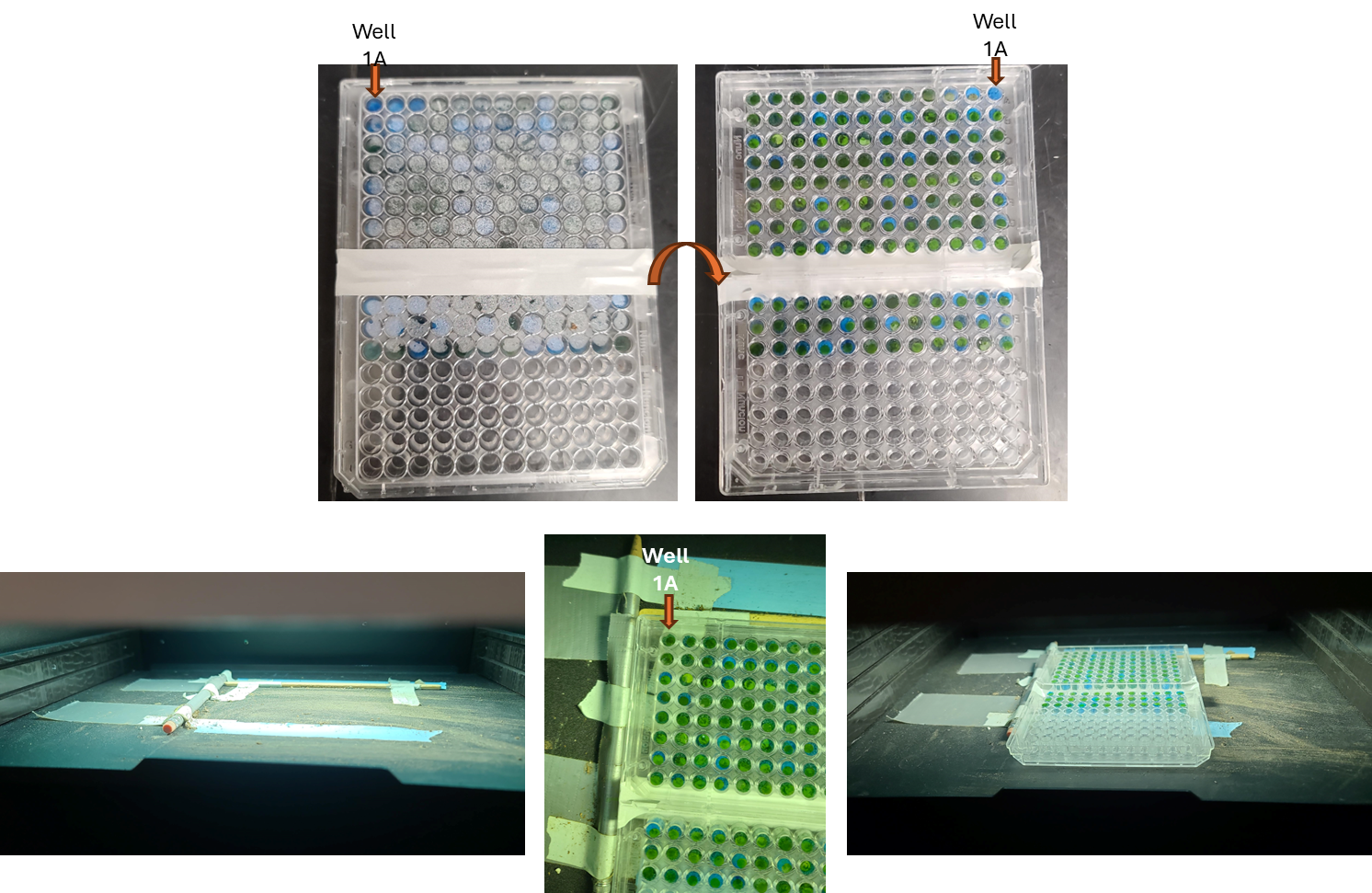
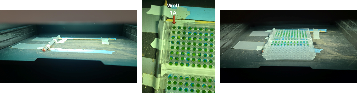
Identify the top left corner of the plate containing well 1A. Typically the 96 well plates have two corners that are truncated and two that are full corners to help orient the plate, even in the dark.

Flip the plate on the vertical edge so the adaxial surface of the plated leaf discs faces up. Well 1A should now be in the top right corner.
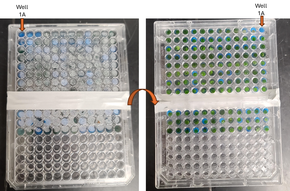
Place the plate on the shelf with the top left corner of the plate snugly against the guardrails for the field of view.
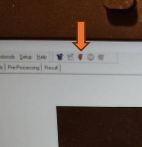
Select the lightening bolt icon to begin analysis.
Progress and time to run completion will be displayed on the bottom of the live screen.

At the completion of the analysis, the Pre-Processing window should automatically open.
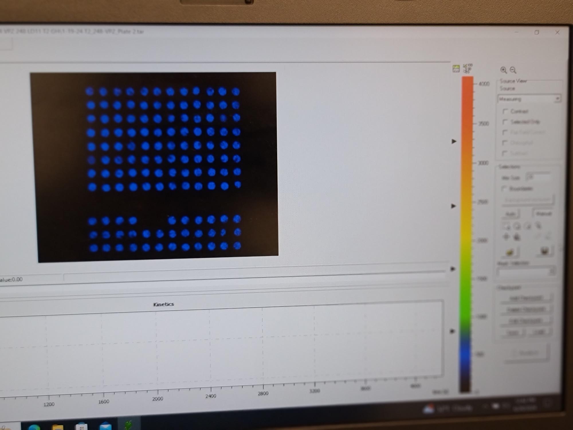
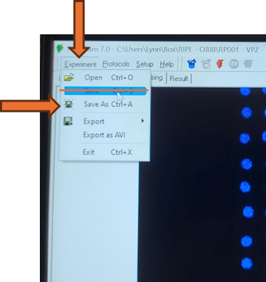
The Fluorcam is not configured with an auto-save. Save the complete experiment .tar file by selecting Experiment-> Save from the top menu bar.
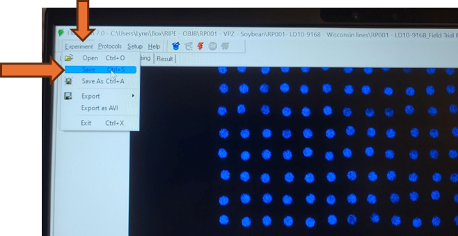
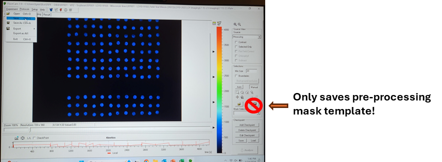
Do not use the save icon on the Pre-Processing window. The save icon on the Pre-Processing window will only save a pre-processing mask and all run data will be lost.
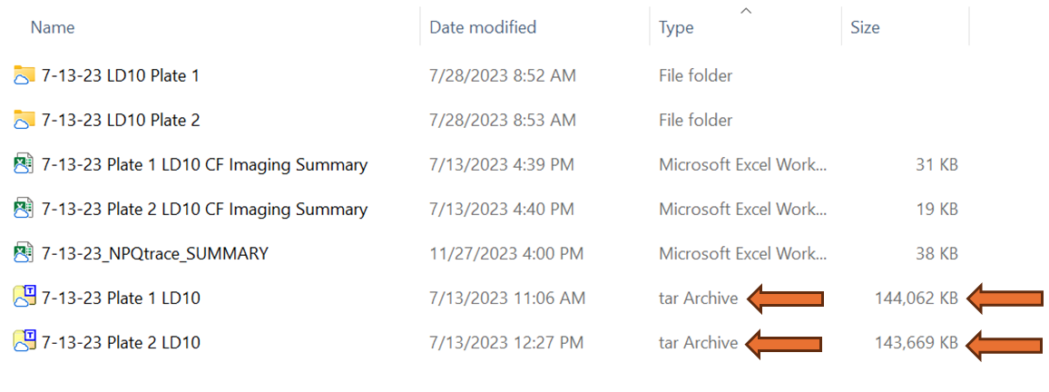
Verify that the file was saved correctly by navigating to the PC location where the analysis file was saved. The file should appear as file type "tar Archive" and be approximately 140,000 KB file size if the provided NPQ protocol was used. This tar file contains the size calibration, protocols, and all analysis imaging data. If a mask was applied or result analysis was initiated it will also contain that data but these two actions can also be performed at a later time.
Remove the completed sample plate from the imager and confirm that well 1A was in the top right corner and well 12A was in the top left corner and that the file name matches the plate analyzed.
Analyze the next plate or sample set following the same steps. The software does not need to be cleared. Once the next plate is ready for analysis, select the lightening bolt icon to begin. Be aware that as soon as the new analysis has begun, the previous pre-processing and results will not be accessible from the file being live generated. When this analysis is complete, be sure to use "save as", not save, to avoid overwriting the previous analysis. For added security to avoid overwriting analysis runs, the software can be closed completely but the size calibration and loading of the protocol will have to be repeated every time the software is closed and then opened.