Minimally invasive longitudinal intravital imaging of cellular dynamics in intact long bone
Nayan Deger Bhattacharyya, Wunna Kyaw, Michelle M. McDonald, Rama Dhenni, Abigail K. Grootveld, Ya Xiao, Ryan Chai, Weng Hua Khoo, Linda C. Danserau, C. Marcelo Sergio, Paul Timpson, Woei Ming Lee, Peter I. Croucher, Tri Giang Phan
intravital imaging
osteoclasts
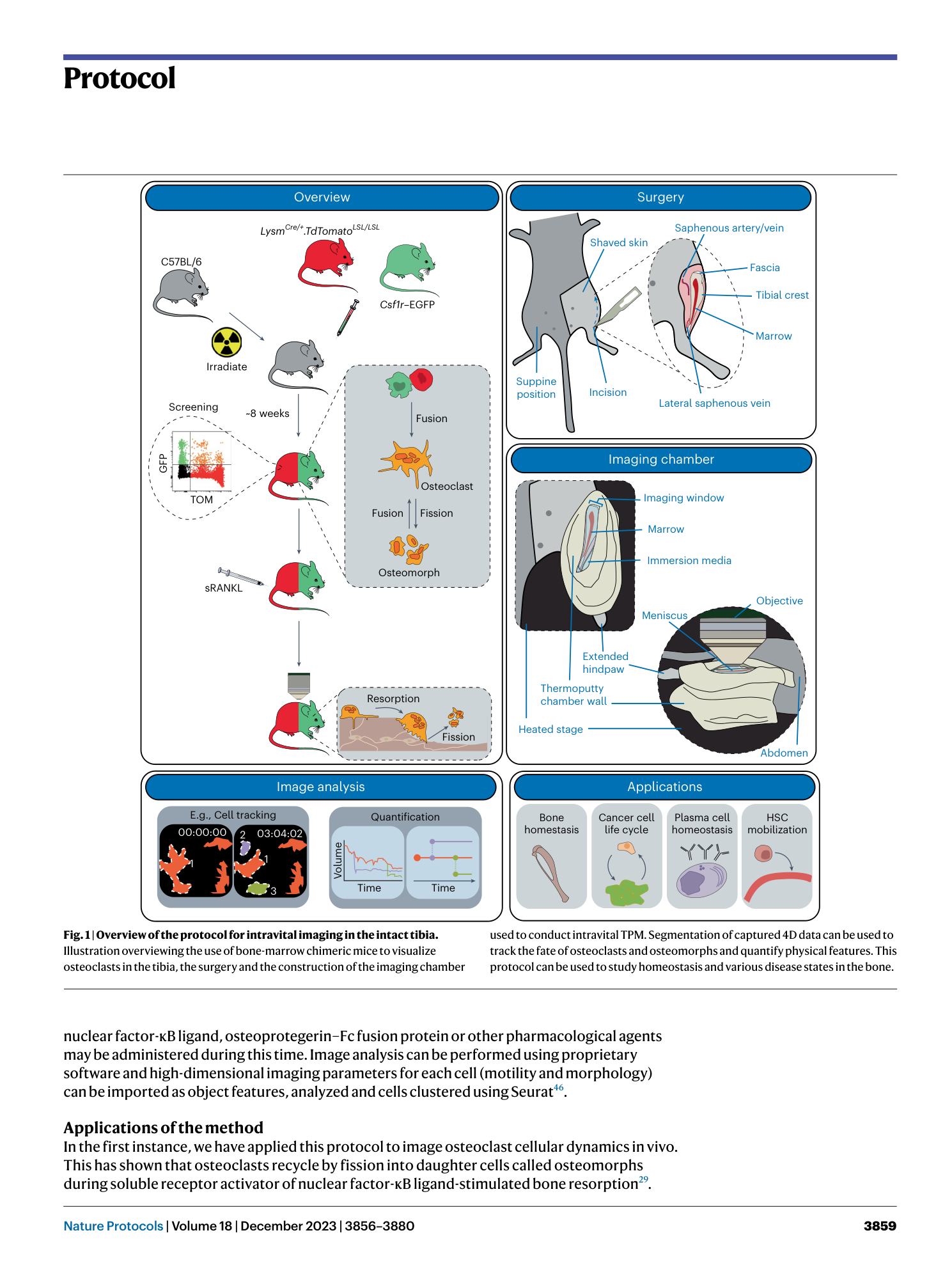
bone marrow chimeras
longitudinal monitoring
minimally invasive surgery


Supplementary information
Reporting Summary
Supplementary Video 1
Intravital imaging of steady-state osteoclast recycling related to Fig. 3. a , 3D rotation of the tibial bone (SHG, blue) surface. b , Video depicting osteoclast fission and fusion. Arrow denotes a fission and fusion event in an GFP+ (CSF1R, green) TOM+ (LysM, red) osteoclasts. c , Video depicting the cell fate mapping of segmented osteoclast surfaces, as denoted by the underground plots (UG) in Fig. 3b
Supplementary Video 2
Intravital imaging of steady-state osteoclast recycling in the proximal site of the tibia, related to the top of Fig. 4a,b. a , 3D rotation of the tibial bone (SHG, blue) surface and fused GFP+ (CSF1R, green), TOM+ (LysM, red) osteoclasts. b , Video depicting slower rates of osteoclast fission and fusion. c , Video depicting the cell fate mapping of segmented osteoclast surfaces, as denoted by the underground plots in
Supplementary Video 3
Intravital imaging of steady-state osteoclast recycling in the distal site of the tibia, related to the bottom of Fig. 4 a,b. a , 3D rotation of the tibial bone (SHG, blue) surface and fused GFP+ (CSF1R, green), TOM+ (LysM, red) osteoclasts. b , Video depicting accelerated rates of osteoclast fission and fusion. c , Video depicting the cell fate mapping of segmented osteoclast surfaces, as denoted by the underground plots in
