Laboratory Maintenance of the Chytrid Fungus Batrachochytrium dendrobatidis
Lillian K. Fritz-Laylin, Lillian K. Fritz-Laylin, Sarah M. Prostak, Sarah M. Prostak
Abstract
The chytrid fungus Batrachochytrium dendrobatidis (Bd) is a causative agent of chytridiomycosis, a skin disease associated with amphibian population declines around the world. Despite the major impact Bd is having on global ecosystems, much of Bd ’s basic biology remains unstudied. In addition to revealing mechanisms driving the spread of chytridiomycosis, studying Bd can shed light on the evolution of key fungal traits because chytrid fungi, including Bd , diverged before the radiation of the Dikaryotic fungi (multicellular fungi and yeast). Studying Bd in the laboratory is, therefore, of growing interest to a wide range of scientists, ranging from herpetologists and disease ecologists to molecular, cell, and evolutionary biologists. This protocol describes how to maintain developmentally synchronized liquid cultures of Bd for use in the laboratory, how to grow Bd on solid media, as well as cryopreservation and revival of frozen stocks. © 2021 The Authors. Current Protocols published by Wiley Periodicals LLC.
Basic Protocol 1 : Reviving cryopreserved Bd cultures
Basic Protocol 2 : Establishing synchronized liquid cultures of Bd
Basic Protocol 3 : Regular maintenance of synchronous Bd in liquid culture
Alternate Protocol 1 : Regular maintenance of asynchronous Bd in liquid culture
Basic Protocol 4 : Regular maintenance of synchronous Bd on solid medium
Alternate Protocol 2 : Starting a culture on solid medium from a liquid culture
Basic Protocol 5 : Cryopreservation of Bd
INTRODUCTION
The chytrid fungus Batrachochytrium dendrobatidis (Bd) is a causative agent of chytridiomycosis, a skin disease that is associated with significant population declines of amphibians worldwide (O'Hanlon et al., 2018). Bd is one of more than a thousand species of chytrid fungus (James et al., 2006), which are characterized by motile life stages called zoospores. The phylogenetic position of chytrid fungi makes this group, including Bd , well suited for studying the evolution of key cellular traits in the fungal lineage (Medina, Turner, Gordân, Skotheim, & Buchler, 2016; Prostak, Robinson, Titus, & Fritz-Laylin, 2021). For these reasons, there is growing interest in using Bd in the laboratory to answer both ecological and evolutionary questions (Medina & Buchler, 2020). Maintenance of a healthy Bd cell culture is critical for ensuring reproducible results and reducing experimental variability from experiment to experiment and lab to lab. Here, we focus on the two most commonly used methods of culturing Bd —growth in liquid tryptone medium and on tryptone plates. Initial cultures can be obtained from the Collection of Zoosporic Eufungi at the University of Michigan in Ann Arbor, Michigan, USA (https://czeum.herb.lsa.umich.edu/).
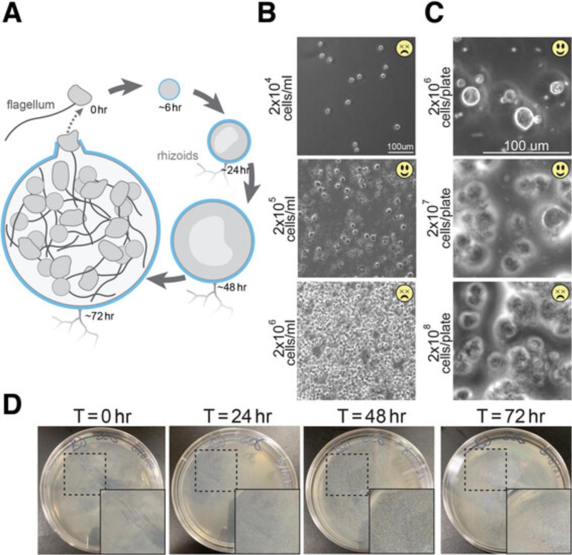
Like all chytrid fungi, Bd has a biphasic life cycle that incorporates both dispersal and growth forms. The life cycle begins with the dispersal form, a motile, flagellated “zoospore” that lacks a cell wall. The zoospore eventually undergoes encystation, the irreversible transition into the growth form: A sessile sporangium that grows a cell wall and hyphal-like rhizoids (Berger, Hyatt, Speare, & Longcore, 2005; see Fig. 1). Sporangia grow and eventually release dozens of new zoospores into the environment to complete the life cycle.

Because zoospores and sporangia each lend themselves to distinct types of experiments, we find it useful to grow cultures that are undergoing synchronous development. For example, while zoospores are used to investigate cell motility and host identification strategies (Moss, Reddy, Dortaj, & San Francisco, 2008; Prostak et al., 2021), sporangia are used to investigate cell wall assembly and composition, as well as cell division (Medina & Buchler, 2020; Prostak et al., 2021). In the laboratory, we grow cells in tissue culture treated (TC-treated) flasks on which Bd sporangia attach. This allows for separation of zoospores from sporangia simply by pouring off the medium. Seeding fresh flasks with these zoospores results in semi-synchronization of the cultures. Tighter synchrony of a population of Bd can be easily attained by thoroughly washing out existing zoospores, adding fresh medium, and collecting zoospores that are released in the following 2 hr. Harvesting the resulting zoospores results in a uniformly age-matched population of cells for downstream developmental studies. Non-TC-treated flasks can be used when growing cultures for cryopreservation, for harvesting sporangia for experimental purposes, or for growth of mixed age (non-synchronized) cultures.
Because Bd cultures are density sensitive, seeding the appropriate number of cells is essential for robust growth of cultures. We recommend using a hemocytometer according to the manufacturer's instructions to accurately count cell density for seeding. See the Internet Resources section for more information on counting cells with the hemocytometer.
CAUTION: Bd is a pathogen of amphibians. Consult with your institution's office of Environmental Health and Safety and follow appropriate precautions to prevent environmental release.
STRATEGIC PLANNING
After thawing, Bd can take up to 1 week to become active enough to begin synchronizing cultures. Once a synchronized culture is established by regular subculturing every 3 days, ensure that the culture is transferred at an appropriate time period to allow cells to grow to the desired developmental stage. For example, if an experiment requires a pure culture of zoospores, start the culture 72-hr prior to when the cells are needed. For germlings, start the culture 24-hr prior, and for mid-stage sporangia, 48-hr. To have healthy cells available daily, it is ideal to have three active cultures at any time, staggering their life cycles such that each culture is transferred to new medium on its own day (Supplementary Fig. 1).
Basic Protocol 1: REVIVING CRYOPRESERVED Bd CULTURES
This protocol outlines the method for thawing Bd from frozen stocks to have cultures in the lab. All steps except for centrifugation should be performed in a biosafety cabinet to both contain and maintain the sterility of the culture. Be sure to spray items with 70% ethanol before placing into the biosafety cabinet to ensure sterility of the culture is maintained.
Materials
-
Frozen Bd JEL423 stock (frozen in 10% DMSO; initial cultures from Collection of Zoosporic Eufungi, University of Michigan: https://czeum.herb.lsa.umich.edu/)
-
1% tryptone, sterile (MilliporeSigma, T7293)
-
70% ethanol solution (for decontaminating supplies and flow hood)
-
50-ml conical tubes, sterile (Eppendorf, 0030122178)
-
Laminar flow hood (BSC Class II or equivalent)
-
Tissue culture microscope (such as Nikon Eclipse Ts2)
-
Centrifuge and adaptors for use with 50-ml tubes (such as Centrifuge 5415R, Eppendorf, 5401000137)
-
Tissue culture treated culture flasks with vent cap, sterile (Cell Treat, 229331, 229341, or 229351)
-
1.5-ml centrifuge tubes, sterile (Eppendorf, 022363204)
-
20- to 200-μl micropipet (such as Eppendorf, 3123000055)
-
2- to 20-μl micropipet (such as Eppendorf, 3123000039)
-
Filter tips for micropipets, sterile (such as USA Scientific, 1122-1830)
-
Pipet controller (Drummond Scientific, 4-000-100 or equivalent)
-
Serological pipets, sterile
-
Neubauer-improved hemocytometer (such as Thermo Fisher Scientific, 02-671-51B)
NOTE : All solutions and equipment coming into contact with cells must be sterile. Appropriate sterile technique and biosafety flow hood should be used.
1.Thaw 1 ml frozen Bd by swirling tube in a lukewarm water bath.
2.Spray tube and other necessary items with 70% ethanol and place in BSC.
3.Use a pipet to add contents of tube to 9 ml 1% tryptone in a 50-ml conical tube.
4.Rinse cryotube with an additional 1 ml 1% tryptone to gather any remaining cells, adding this to the volume from step 2.
5.Spin cells down at 2,500 × g for 5 min to separate DMSO-containing medium from cells.
6.Pour off supernatant and resuspend cells in 10 ml 1% tryptone.
7.Prepare a 1:10 dilution (10 μl cells, 90 μl medium) of zoospores from the resuspended Bd into a 1.5-ml centrifuge tube.
8.Count the number of cells in the diluted aliquot using a hemocytometer and determine the density of resuspended cells in the conical tube.
9.Add appropriate volumes of resuspended cells and new medium into a 25-cm2 TC-treated culture flask at a final concentration of 2 × 10⁵ cells/ml in 10 ml.
10.Check under microscope daily to assess activity level.
Basic Protocol 2: ESTABLISHING SYNCHRONIZED LIQUID CULTURES OF Bd
It is often advantageous to synchronize cultures to ensure that cells harvested for experiments are at a similar stage of development. This helps to minimize variability within a sample. This protocol outlines how to obtain a synchronized culture of Bd in liquid medium. All steps except for centrifugation should be performed in a biosafety cabinet to both contain and maintain the sterility of the culture. Be sure to spray items with 70% ethanol before placing into the biosafety cabinet to ensure sterility of the culture is maintained.
Materials
-
Active, asynchronous Bd JEL423 culture (Collection of Zoosporic Eufungi, University of Michigan: https://czeum.herb.lsa.umich.edu/)
-
1% tryptone, sterile (MilliporeSigma, T7293)
-
70% ethanol solution (for decontaminating supplies and flow hood)
-
50-ml conical tubes, sterile (Eppendorf, 0030122178)
-
Laminar flow hood (BSC Class II or equivalent)
-
Tissue culture microscope (such as Nikon Eclipse Ts2)
-
Centrifuge and adaptors for use with 50-ml tubes (such as Centrifuge 5415R, Eppendorf, 5401000137)
-
Tissue culture treated culture flasks with vent cap, sterile (Cell Treat, 229331, 229341, or 229351)
-
Steriflip filters, 20 μm (MilliporeSigma, SCNY00020)
-
1.5-ml centrifuge tubes, sterile (Eppendorf, 022363204)
-
20- to 200-μl micropipet (such as Eppendorf, 3123000055)
-
2- to 20-μl micropipet (such as Eppendorf, 3123000039)
-
Filter tips for the micropipets, sterile (such as USA Scientific, 1122-1830)
-
Pipet controller (Drummond Scientific, 4-000-100 or equivalent)
-
Serological pipets, sterile
-
Neubauer-improved hemocytometer (such as Thermo Fisher Scientific, 02-671-51B)
NOTE : All solutions and equipment coming into contact with cells must be sterile. Appropriate sterile technique and biosafety flow hood should be used.
1.To achieve a synchronous culture, filter the asynchronous culture through a 20-μm Steriflip filter into a 50-ml conical tube.
2.Prepare a 1:10 dilution (10 μl cells, 90 μl medium) of the filtered zoospores into a 1.5-ml centrifuge tube.
3.Add appropriate volumes of filtered cells and new medium into a 25-cm2 TC-treated culture flask at a final concentration of 2 × 10⁵ cells/ml in 10 ml.
4.Incubate flask at 24˚C, subculturing to a new flask every 3 days (see Basic Protocol 3).
Basic Protocol 3: REGULAR MAINTENANCE OF SYNCHRONOUS Bd IN LIQUID CULTURE
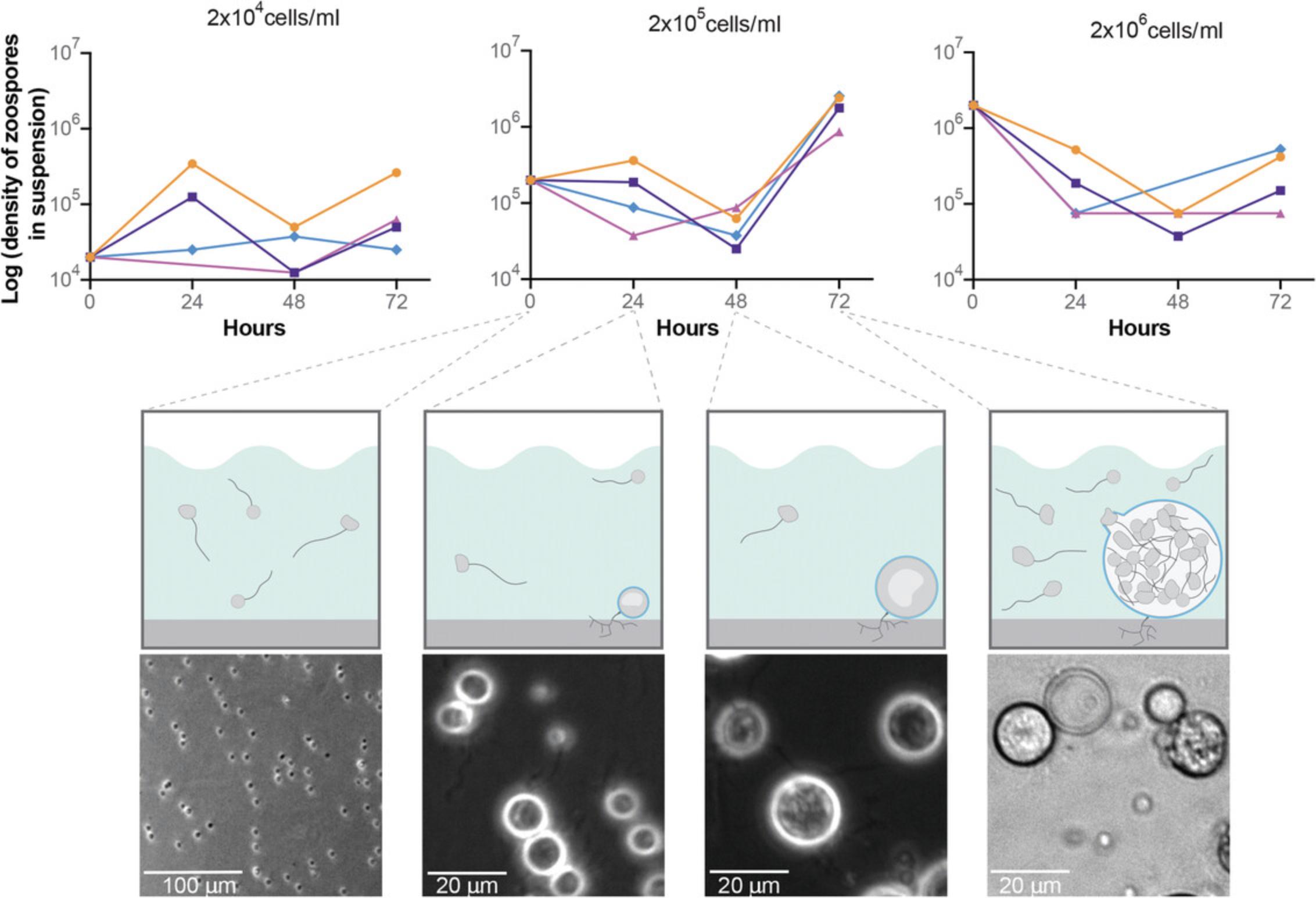
This protocol outlines how to maintain a synchronized culture of Bd in liquid medium. It is essential to seed Bd at the appropriate density: If the culture is too sparse, sporangia may fail to release zoospores; if it is too dense, cells will fail to develop (Fig. 2). We usually seed cells in flasks at ∼2 × 10⁵ cells/ml with a final volume of 10 ml for a 25-cm2 flask; 25 ml for a 75-cm2 flask; and 60 ml for a 175-cm2 flask. Note that because Bd is a deadly pathogen to amphibians we typically treat it as a BSL-II organism and conduct cell culture in a laminar flow biosafety cabinet. We also autoclave or treat with freshly diluted 10% bleach all items that come in contact with cultures, including tissue culture plastics.

Materials
-
Active, synchronous culture of Bd JEL423 in liquid medium (see Basic Protocol 1 and Basic Protocol 2)
-
Tryptone liquid medium, sterile (1%, w/v, MilliporeSigma, T7293; see recipe)
-
70% ethanol solution (for decontaminating supplies and flow hood)
-
Laminar flow hood (BSC Class II or equivalent)
-
Tissue culture microscope (such as Nikon Eclipse Ts2)
-
TC-treated culture flasks with vent cap, sterile (Cell Treat, 229331, 229341, or 229351)
-
1.5-ml centrifuge tubes, sterile (Eppendorf, 022363204)
-
20- to 200-μl micropipet (such as Eppendorf, 3123000055)
-
2- to 20-μl micropipet (such as Eppendorf, 3123000039)
-
Filter tips for the micropipets, sterile (such as USA Scientific, 1122-1830)
-
Pipet controller (Drummond Scientific, 4-000-100 or equivalent)
-
Serological pipets, sterile
-
Neubauer-improved hemocytometer (such as Thermo Fisher Scientific, 02-671-51B)
-
Centrifuge and adaptors for use with 50-ml tubes (such as Centrifuge 5415R, Eppendorf, 5401000137)
NOTE : All solutions and equipment coming into contact with cells must be sterile. Appropriate sterile technique and biosafety flow hood should be used.
1.After thawing Bd (see Basic Protocol 1) and synchronizing the culture (see Basic Protocol 2), transfer (or “subculture”) the cells to new medium every 3 days as follows.
2.Prepare a 1:10 dilution of zoospores in suspension from a 3-day-old flask of Bd into a 1.5-ml centrifuge tube by removing 10 μl medium from the mature culture flask and adding to 90 μl medium in a 1.5-ml centrifuge tube. Gently flick tube to mix.
3.Count cell density in the diluted aliquot using a hemocytometer; determine the density of cells in the flask using the appropriate formula.
4.Add appropriate volumes of cells from the 3-day-old flask and new medium to subculture into a new flask at a final concentration of 2 × 10⁵ cells/ml.
5.Close the cap of the flask, gently lay it on its flat side, and grow at 24˚C for 3 days.
Alternate Protocol 1: REGULAR MAINTENANCE OF ASYNCHRONOUS Bd IN LIQUID CULTURE
Bd can be cultured asynchronously, though the culture will be less predictable. Maintaining an asynchronous culture requires daily observation of flasks and is difficult to determine cell density. Subculturing would take place as needed or when the flask becomes too dense by eye.
Additional Materials (see also Basic Protocol 1)
- Non-TC-treated culture flasks with vent cap, sterile (Cell Treat, 229500, 229510, or 229520)
1.After thawing Bd into a non-TC-treated flask (see Basic Protocol 1), transfer (or “subculture”) the cells to new medium as needed, as follows.
2.Prepare a 1:10 and 1:20 dilution of the mature cells into new medium in a non-TC-treated flask.
3.Close the cap of the flasks, gently lay on their flat sides, and grow at 24˚C until culture becomes too dense and needs to be sub-cultured again.
Basic Protocol 4: REGULAR MAINTENANCE OF SYNCHRONOUS Bd ON SOLID MEDIUM
This protocol outlines how to maintain Bd on agar plates, which can be helpful for isolation of individual colonies. All steps except for centrifugation should be performed in a biosafety cabinet to both contain and maintain the sterility of the culture. Be sure to spray items with 70% ethanol before placing into the biosafety cabinet to ensure sterility of the culture is maintained.
Materials
-
Active, healthy culture of Bd JEL423 (Collection of Zoosporic Eufungi, University of Michigan: https://czeum.herb.lsa.umich.edu/)
-
Solid medium (1% tryptone, MilliporeSigma, T7293; 1.5% agar, Thermo Fisher Scientific, BP1423)
-
Dilute Salts (DS) solution (see recipe)
-
Laminar flow hood (BSC Class II or equivalent)
-
Tissue culture microscope (such as Nikon Eclipse Ts2)
-
Neubauer-improved hemocytometer (such as Thermo Fisher Scientific, 02-671-51B)
-
1.5-ml centrifuge tubes, sterile (Eppendorf, 022363204)
-
1,000-μl micropipet (such as Eppendorf, 3124000121)
-
20- to 200-μl micropipet (such as Eppendorf, 3123000055)
-
2- to 20-μl micropipet (such as Eppendorf, 3123000039)
-
Filter tips for micropipets, sterile (such as USA Scientific, 1122-1830)
-
Glass beads, sterile (for spreading)
NOTE : All solutions and equipment coming into contact with cells must be sterile. Appropriate sterile technique and biosafety flow hood should be used.
1.Collect zoospores from a plate of actively growing Bd.
2.Take 10 μl of the liquid and count zoospores using a hemocytometer; determine the density of cells in the flask using the appropriate formula.
3.Dilute cells to a final concentration between 2 × 10⁶ and 2 × 10⁷ cells/ml in 1 ml DS.
4.Add 1 ml diluted cells to a new 1% tryptone plate.
5.Gently shake the new plate to spread the liquid around the agar.
6.Incubate plates at 24˚C.
7.Transfer cells to new plates every 3 days to keep the culture healthy.
Alternate Protocol 2: STARTING A CULTURE ON SOLID MEDIUM FROM A LIQUID CULTURE
Although preparing solid medium is more time consuming than preparing liquid medium, growing Bd on plates has several advantages, for example, easier detection of a contaminated culture. (See Commentary, Background Information for more detail.)
Additional Materials (see also Basic Protocol 3)
- Active, healthy liquid culture of Bd JEL423 (Collection of Zoosporic Eufungi, University of Michigan: https://czeum.herb.lsa.umich.edu/)
1.Prepare a 1:10 dilution (10 μl cells, 90 μl medium) of zoospores from a 3-day flask into a 1.5-ml centrifuge tube.
2.Count zoospores from diluted aliquot using a hemocytometer; determine density of cells in the 3-day-old flask.
3.Dilute cells to between 2 × 10⁶ and 2 × 10⁷ cells/ml in 1 ml DS.
4.Add 1 ml diluted cells to a new 1% tryptone plate.
5.Gently shake the new plate to spread the liquid around the agar.
6.Incubate plates at 24˚C.
7.Check on plates daily to assess activity levels, ensuring zoospores are present and mobile.
8.Transfer cells to new plates every 3 days to keep the culture healthy.
Basic Protocol 5: CRYOPRESERVATION OF Bd
The following method is adapted from Boyle et al. (2003), which involves slow freezing of ampules of cells resuspended in a cryopreservant and long-term storage in liquid nitrogen or at -80°C. All steps except for centrifugation should be performed in a biosafety cabinet to both contain and maintain the sterility of the culture. Be sure to spray items with 70% ethanol before placing into the biosafety cabinet to ensure sterility of the culture is maintained. We recommend cryopreserving multiple vials in parallel and testing the viability of the cultures by reviving one vial after at least 2 weeks of cryostorage.
Materials
-
Active, healthy culture of Bd JEL423 (Collection of Zoosporic Eufungi, University of Michigan: https://czeum.herb.lsa.umich.edu/) grown in a non-TC-treated flask
-
Cryopreservation medium: Sterile 1% tryptone (MilliporeSigma, T7293), 10% sterile DMSO (MilliporeSigma, D2650)
-
70% ethanol solution (for decontaminating supplies and flow hood)
-
Laminar flow hood (BSC Class II or equivalent)
-
Tissue culture microscope (such as Nikon Eclipse Ts2)
-
50-ml conical tubes, sterile (Eppendorf, 0030122178)
-
Centrifuge and adaptors for use with 50-ml tubes (such as Centrifuge 5415R, Eppendorf, 5401000137)
-
Cryovials with rubber gaskets, sterile (Corning, 431416)
-
Mr. Frosty isopropanol container (Thermo Fisher Scientific, 5100-0001)
-
Neubauer-improved hemocytometer
-
-80°C freezer
-
Liquid nitrogen freezer
NOTE : All solutions and equipment coming into contact with cells must be sterile. Appropriate sterile technique and biosafety flow hood should be used.
1.Remove cells from culture flask and transfer to a 50-ml conical centrifuge tube.
2.Centrifuge cells at 2,500 × g for 5 min; remove supernatant.
3.Resuspend cells in 5 ml cryopreservation medium pre-chilled to 4°C.
4.Aliquot 1 ml cells into labeled cryovials.
5.Transfer vials to pre-chilled (4°C) Mr. Frosty isopropanol container and store container in -80°C freezer.
REAGENTS AND SOLUTIONS
Dilute salts (DS) solution, 1×
- 500 μl DS stock solution I (see recipe)
- 100 μl DS stock solution II (see recipe)
- 1 L sterile water
- Prepare solution in a sterile laminar flow hood with sterile supplies.
- Store at room temperature for up to 12 months.
From: Machlis (1958).
Dilute salts (DS) stock solution I, 10×
- 68.05 g potassium phosphate monobasic (KH2PO4; MilliporeSigma, P0662)
- 87.09 g potassium phosphate dibasic (K2HPO4; MilliporeSigma, P3786)
- 66.04 g ammonium phosphate dibasic, ((NH4)2HPO4; MilliporeSigma, 215996)
- 500 ml water
- Sterilize by filtration.
- Store at room temperature for up to 12 months.
Final concentrations: 0.5 mM KH2PO4; 0.5 mM K2HPO4; 0.5 mM (NH4)2HPO4.
Dilute salts (DS) stock solution II, 10×
- 25.42 g magnesium chloride (MgCl2; MilliporeSigma, M8266)
- 18.38 g calcium chloride dihydrate (CaCl2; MilliporeSigma, C1016)
- 250 ml water
- Sterilize by filtration.
- Store at room temperature for up to 12 months.
Final concentrations: 0.05 mM MgCl2; 0.05 mM CaCl2.
Tryptone liquid medium
- 10 g tryptone (1% w/v; MilliporeSigma, T7293)
- 1 L water
- For plates: Add 15 g agar (1.5%, w/v, Thermo Fisher Scientific, BP1423)
- Sterilize by autoclaving or by filtration.
- Store liquid at room temperature and plates at 4°C for up to 6 months.
COMMENTARY
Background Information
Although chytrids have been grown in the laboratory since the mid-1900s, Bd was first described in 1999 by Joyce Longcore and colleagues (Longcore, Pessier, & Nichols, 1999). This initial publication described the isolation of Bd from infected frog skin and growth on solid PmTG medium (1 g peptonized milk, 1 g tryptone, 5 g glucose, 10 g agar, 1 L distilled water, with 400 mg streptomycin sulfate and 200 mg penicillin-G added after autoclaving) at temperatures ranging from 20°C to 23°C. Week-old sporangia from these isolation plates were transferred to TGhL agar (16 g tryptone, 4 g gelatin hydrolysate, 2 g lactose, 12 g agar, 1 L distilled water). Since that time, most laboratories have adopted 1% tryptone as the primary growth medium.
There are advantages and disadvantages to growing Bd in liquid media. An obvious advantage is the ease at which one can scale up the culture simply by using different sized flasks. Growing Bd in liquid also facilitates synchronization of the culture, allowing one to harvest cells of the same stage of development. However, using synchronized liquid cultures requires the maintenance of multiple cultures to ensure that zoospores will be available for daily use. We routinely split each culture every 3 days in rotation, meaning that one of the three must be split every day, including weekends (Supporting Information Fig. 1). However, flasks of Bd can be left at 4°C for at least 1 month if not being actively used in the laboratory and the culture revived by subculturing when needed.
We more often grow Bd liquid media because preparing solid media is more time consuming than liquid, and cells may take longer to recover from overgrowth when grown on plates than in liquid media or when revived from cryopreservation. However, there are times at which growing the organism on solid media is preferable. For example, growing Bd on plates allows for easy detection of contamination in the culture, as low levels of bacteria are readily visible as colonies when the culture is grown at low density on solid medium. Growing Bd on plates also allows for the selection of individual colonies for isolating single cell clones.
Critical Parameters
The most sensitive aspect of this protocol is cell density and contamination. Bd does not grow well at very low or very high densities. Maintaining a sterile, pure culture of Bd is also imperative for gathering trusted results from your experiments.
Troubleshooting
See Table 1 for troubleshooting recommendations.
| Problem | Possible cause | Solution |
|---|---|---|
| Little or no zoospores released when grown in liquid culture | Density too low | Make sure the final density of cells in the new flask is 2 × 10⁵ cells/ml |
| Little or no zoospores released when grown in liquid culture | Density too high | Make sure the final density of cells in the new flask is 2 × 10⁵ cells/ml |
| Contamination during regular subculturing | Improper sterile technique or contaminated medium or supplies | Properly dispose of all active cultures, restart cultures from frozen stocks, using all new, sterile medium and supplies |
| Contamination after thawing from frozen stocks | Contaminated stock cultures |
Properly dispose of all active cultures; thaw another stock, and if cultures are still contaminated, order new cells Freeze new stocks and restart cultures from these new stocks; once new cultures are active, properly dispose of all old frozen stocks to ensure they are not used in the future |
| No growth after thawing | Frozen stock cells are dead | Sometimes individual vials do not survive cryopreservation. Other times the entire batch of frozen cells are nonviable. If no cells have grown 1 week after thawing a stock, try thawing another vial from the same batch. If that also does not grow, throw away all vials from the bad batch to ensure you don't use them in the future. |
| A lot of floating sporangia in liquid culture | Flasks are not tissue culture treated |
Ensure you are using tissue culture treated culture flasks when growing daily-use cultures; large amounts of clumps of cells may interfere with synchrony The best brand of culture flasks to use with Bd are CellTreat flasks (Cell Treat, 229331, 229341, or 229351) |
| A lot of floating sporangia in liquid culture | Zoospores preferentially adhering to sporangia instead of flasks |
If many clumps persist in CellTreat flasks the problem may be zoospores sticking to sporangia; clumps of cells may interfere with synchrony To remove clumps, filter liquid culture through a 20-μm Steriflip filter (MilliporeSigma, SCNY00020) to ensure only zoospores are in suspension before transferring cells to a new flask |
Understanding Results
Bd cultures are healthiest when transferred at medium densities, ∼2 × 10⁵ cells/ml or between 2 × 10⁶ and 2 × 10⁷ cells/plate. To gauge whether a synchronous Bd culture is healthy, check to see that the majority of zoospores are released, ∼72-hr after transferring cells to new medium (Fig. 2). The amount of zoospores released after this time is typically an order of magnitude higher than the density at which the culture was transferred. There should also be space between sporangia (Fig. 1B), though some clumps may form.
Time Considerations
The process of transferring cultures depends on how many flasks or plates are being made and typically takes between 15 and 45 min. Cryopreserving cultures again depends on the number of vials being frozen but typically takes ∼30 min to prepare and 24 to 48 hr to completely freeze. Thawing cultures typically takes 15 min and usually 1 week until the cells are at proper density and running on a synchronous, 3-day life cycle.
Acknowledgments
This work was supported by the National Science Foundation under award IOS-1827257 to L.F.-L. The authors thank Dr. Andrew Swafford for feedback on the manuscript.
Author Contributions
Sarah M. Prostak : Conceptualization, data curation, formal analysis, investigation, methodology, visualization, writing original draft, writing review and editing; Lillian K. Fritz-Laylin : Conceptualization, data curation, formal analysis, funding acquisition, investigation, methodology, project administration, supervision, visualization, writing original draft, writing review and editing.
Conflict of Interest
The authors declare no conflict of interest.
Open Research
Data Availability Statement
The data that support this protocol are available in the figures and supplement of this article.
Supporting Information
| Filename | Description |
|---|---|
| cpz1309-sup-0001-FigureS1.eps19.2 MB | Supporting Information Figure 1. Timing of culture maintenance. To ensure zoospores are available for use daily, we recommend maintaining 3 active cultures, splitting one each day on a 3-week rotating schedule. Each color represents an active culture. |
Please note: The publisher is not responsible for the content or functionality of any supporting information supplied by the authors. Any queries (other than missing content) should be directed to the corresponding author for the article.
Literature Cited
- Berger, L., Hyatt, A. D., Speare, R., & Longcore, J. E. (2005). Life cycle stages of the amphibian chytrid Batrachochytrium dendrobatidis. Diseases of Aquatic Organisms , 68(1), 51–63. doi: 10.3354/dao068051.
- Boyle, D. G., Hyatt, A. D., Daszak, P., Berger, L., Longcore, J. E., Porter, D., … Olsen, V. (2003). Cryo-archiving of Batrachochytrium dendrobatidis and other chytridiomycetes. Diseases of Aquatic Organisms , 56(1), 59–64. doi: 10.3354/dao056059.
- James, T. Y., Kauff, F., Schoch, C. L., Matheny, P. B., Hofstetter, V., Cox, C. J., … Vilgalys, R. (2006). Reconstructing the early evolution of fungi using a six-gene phylogeny. Nature , 443(7113), 818–822. doi: 10.1038/nature05110.
- Longcore, J. E., Pessier, A. P., & Nichols, D. K. (1999). Batrachochytrium dendrobatidis gen. et sp. nov., a chytrid pathogenic to amphibians. Mycologia , 91(2), 219. doi: 10.2307/3761366.
- Machlis, L. (1958). Evidence for a sexual hormone in allomyces. Physiologia Plantarum , 11(1), 181–192. doi: 10.1111/j.1399-3054.1958.tb08436.x.
- Medina, E. M., & Buchler, N. E. (2020). Chytrid fungi. Current Biology , 30(10), R516–R520. doi: 10.1016/j.cub.2020.02.076.
- Medina, E. M., Turner, J. J., Gordân, R., Skotheim, J. M., & Buchler, N. E. (2016). Punctuated evolution and transitional hybrid network in an ancestral cell cycle of fungi. eLife , 5, e09492. doi: 10.7554/eLife.09492.
- Moss, A. S., Reddy, N. S., Dortaj, I. M., & San Francisco, M. J. (2008). Chemotaxis of the amphibian pathogen Batrachochytrium dendrobatidis and its response to a variety of attractants. Mycologia , 100(1), 1–5. doi: 10.1080/15572536.2008.11832493.
- O'Hanlon, S. J., Rieux, A., Farrer, R. A., Rosa, G. M., Waldman, B., Bataille, A., … Fisher, M. C. (2018). Recent Asian origin of chytrid fungi causing global amphibian declines. Science , 360(6389), 621–627. doi: 10.1126/science.aar1965.
- Prostak, S. M., Robinson, K. A., Titus, M. A., & Fritz-Laylin, L. K. (2021). The actin networks of chytrid fungi reveal evolutionary loss of cytoskeletal complexity in the fungal kingdom. Current Biology , 31(6), 1192–1205.e6. doi: 10.1016/j.cub.2021.01.001.
Internet Resources
- https://www.hemocytometer.org/using-hemocytometer-to-count-cells-in-6-steps/
- https://www.hemocytometer.org/hemocytometer-protocol/
Guides to counting cells using a Neubauer-improved (NI) hemocytometer. The information outlines how to count different types of cells using an NI hemocytometer.
Citing Literature
Number of times cited according to CrossRef: 3
- Rebecca J. Webb, Alexandra A. Roberts, Catherine Rush, Lee F. Skerratt, Mark L. Tizard, Lee Berger, Small Interfering RNA Mediated Messenger RNA Knockdown in the Amphibian Pathogen Batrachochytrium dendrobatidis, Journal of Basic Microbiology, 10.1002/jobm.202400081, 64 , 8, (2024).
- Rebecca J. Webb, Christopher Cuff, Lee Berger, Glutathione‐Mediated Metal Tolerance in an Amphibian Chytrid Fungus (Batrachochytrium dendrobatidis), Environmental Toxicology and Chemistry, 10.1002/etc.5885, 43 , 7, (1583-1591), (2024).
- Kristyn A. Robinson, Sarah M. Prostak, Evan H. Campbell Grant, Lillian K. Fritz-Laylin, Amphibian mucus triggers a developmental transition in the frog-killing chytrid fungus, Current Biology, 10.1016/j.cub.2022.04.006, 32 , 12, (2765-2771.e4), (2022).

