In vivo identification of astrocyte and neuron subproteomes by proximity-dependent biotinylation
Joselyn S. Soto, Yasaman Jami-Alahmadi, James A. Wohlschlegel, Baljit S. Khakh


Extended
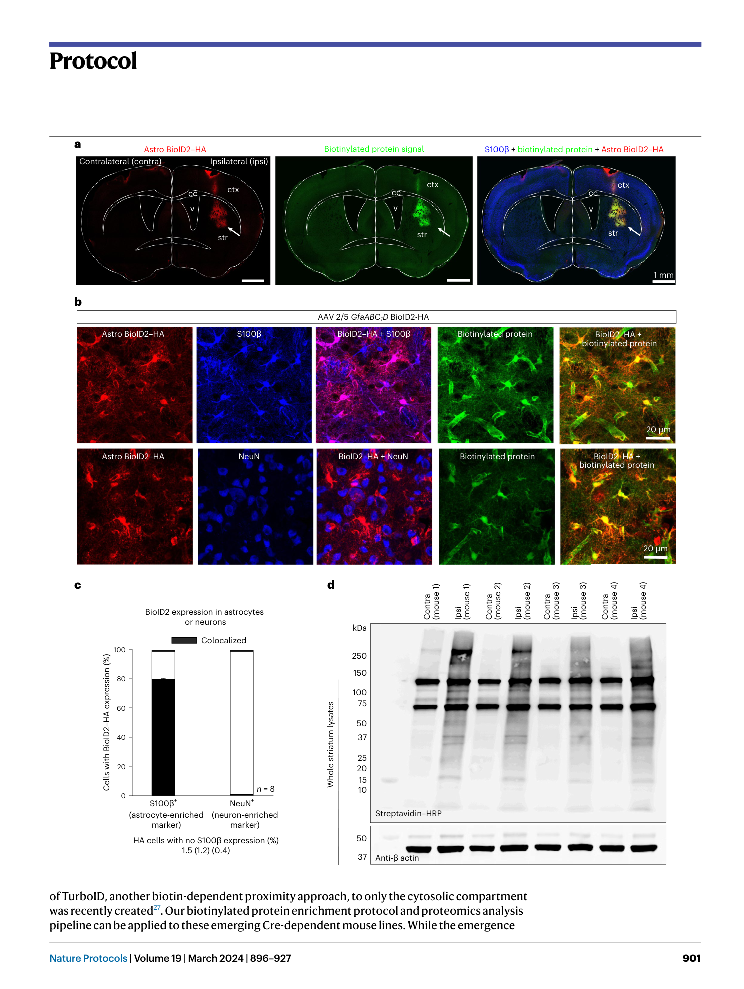
Extended Data Fig. 1 In vivo protein biotinylation in mouse.
a . Photo showing site of subcutaneous biotin injection in mouse for in vivo BioID2 protein biotinylation. b . A successful injection will produce a temporary pocket of biotin under the skin of the mouse (red arrows. The pocket should disappear within 10 minutes.
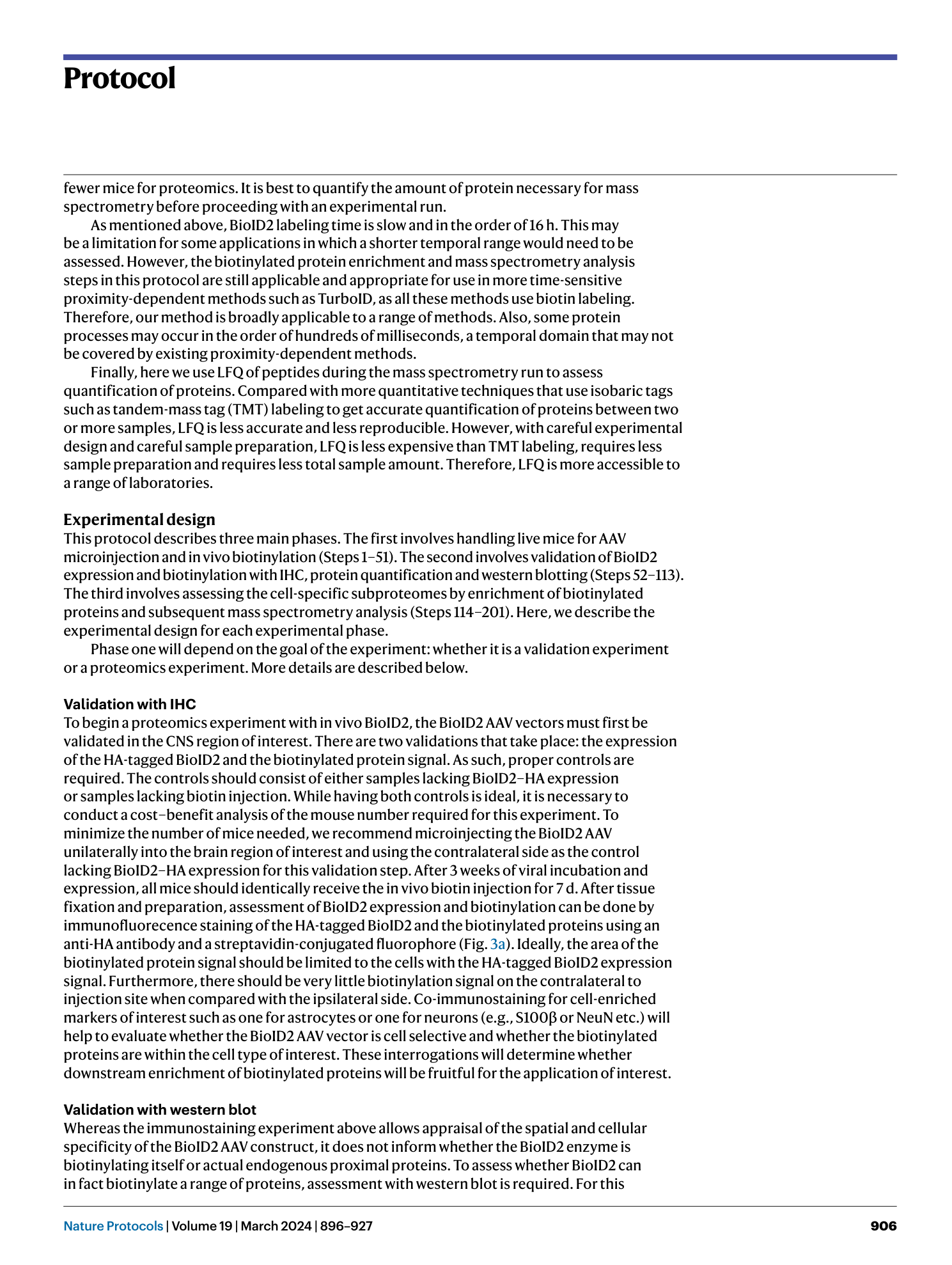
Extended Data Fig. 2 Microdissection setup and tissue homogenization.
a . Photos depicting the microdissection area for removal and isolation of the CNS region of interest. Because the sample must remain cold, the dissection is conducted with a pre-frozen TissueTek cold plate. Standard microdissection tools for striatum are shown. b . Photos of striata in a dounce homogenizer tube that was prefilled with 600 µL of lysis buffer 1 (Step 126) prior to homogenization with pestles. Photo on the right shows homogenate after homogenization with pestles (Step 127).
