Sonication of α-synuclein Fibrils for injection into the mouse brain

Vijay Singh, Marta Castellana-Cruz, Nunilo Cremades, Laura Volpicelli-Daley

Published: 2024-06-17 DOI: 10.17504/protocols.io.14egn337zl5d/v2

Disclaimer

The protocols.io team notes that research involving animals and humans must be conducted according to internationally-accepted standards and should always have prior approval from an Institutional Ethics Committee or Board.

Abstract

Animal models that accurately recapitulate the accumulation of alpha-synuclein (α-syn) inclusions, progressive neurodegeneration of the nigrostriatal system and motor deficits can be useful tools for Parkinson's disease (PD) research. The preformed fibril (PFF) synucleinopathy model in rodents generally displays these PD-relevant features, however, the magnitude and predictability of these events is far from established. We therefore have optimized the sonication protocol of α-syn fibrils to ensure reliable, robust results. The protocol includes steps for re-sonication of PFFs on the day of injection into mice. The sonicated PFFs we received need to be re-sonicated to ensure that any fibrils aggregated due to long-term storage can be optimally fragmented to seed synuclein pathology once injected into mice.

Before start

Sonicating Fibril

Proper sonication is a key step for the fibril model to work. For all in vivo work which involved injecting fibrils into mice or rats, we use the QSonica 700 sonicator with cup horn and tube rack for 1.5 mL polypropylene tubes with a chiller at 16°C. The cup horn sonication produces short fragments which maintain their morphology for 6-8 hours (at least) and can be stored in dry ice overnight, thawed and maintained at room temperature, and therefore remain active after overnight shipments.We found that over time, the heat generated by a probe tip sonicator causes the fibrils to form amorphous aggregates (Figure 1). This is a problem because stereotaxic surgeries can take several hours and the amorphous aggregates that form while the fibrils sit on the bench causes variability and reduces the concentration of seeding competent fragments. Another advantage of using the cup horn sonicator over probe tip is that 25 μL of fibrils can be sonicated, reducing the volume needed. This is also a closed tube system which increases safety. For neuron or cell culture work in which the fibrils are added to media and then the cells immediately after sonication, a probe tip sonicator is okay. Again, this should be performed in a BSL2 hood with all proper PPE (nanoparticle respirator, goggles, gloves etc.). The volume of fibrils to be sonicated cannot be less than 100 μL.

In all cases, we wear PPE when working with fibrils. We clean any spills with 1% SDS.

Figure 1: Transmission electron microscopy of α-synuclein fibrils. Immediately after probe tip or cup horn sonication, long fibrils are broken into small fragments. However, after six hours at room temperature, probe tip sonicated fibrils begin to form amorphous aggregates. With cup horn sonication performed at 16°C, the fragments after 6 hours appear similar in morphology compared to immediately after sonication. When the sonicated fibrils are placed in dry ice overnight, thawed and left at room temperature for 6 hours, the fragments appear similar to immediately after sonication, indicating that overnight shipments will maintain active fragments.

Attachments

Steps

Sonicating Fibrils

1.

WePFFs Fill Qsonica water reservoir with about 900mL.

Note
This protocol is designed to re-sonicate the PFFs that have been sonicated once and stored at -80 degrees Celsius for future injection. On the day of injection, We re-sonicate PFFs to disintegrate any fibrils that aggregated during the storage, ensuring they form optimal fibrils for injection.

2.

Attach cooling system to Qsonica and set the temperature at 10°C.

3.

Thaw and pipet 25-22µL in 1.5mL polystyrene sonication tube.

4.

Place 1.5mL sonication tube with fibrils in Qsonica tube holder.
Note
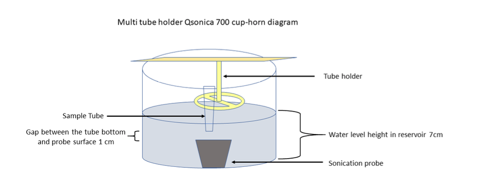
Ensure the reservoir water level is maintained up to the level of the upper meniscus of the PFFs inside the sonication tube.

Figure 2. Diagram of Qsonica700 with multi-tube holder

Note
Sonication cycle parameters: The sonication parameters we are using are as follows: Amplitude at 45%, pulse on and off durations of 3 and 3 seconds respectively, and power for each pulse set at around 110 watts. However, these parameters may not be universally ideal and can vary depending on the specific sonicator used in an individual lab. Therefore, we recommend performing personal optimization of the conditions first. The primary objective is to consistently obtain α-synuclein fragments with an average length of 50 nm. In cases where the fibrils are not adequately fragmented, there might be a low and highly variable production of α-synuclein inclusions.

5.

Total sonication time: 0h 15m 0s.

6.

Sonication pulse on for 3 s and off for 3 s

7.

Amplitude 45%.

Note
After sonication sometimes, there are few droplets inside the sample tube and sometimes not. If droplets are there take out tube and spin it at 1000rpm,°C,0h 0m 10s. Take the sample tube in safety hood and mix the PFF sample with pipette 5 times in and out (avoid introducing bubbles while pipetting).

8.

Sonicate sample for another 7:30 min only if we receive unsonicated PFFs.

Confirming Fragmentation

9.

Note
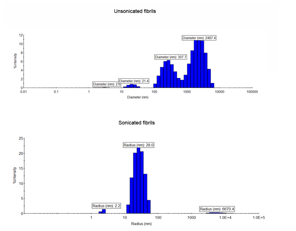
Before injecting fibrils into a large cohort of mice and waiting several months for results, researchers should ensure that their sonication protocol results in sufficient fibril fragmentation. We use a dynamic light scattering detector as a quick and reliable method to check for fragmentation. Transmission electron microscopy is another method that can be used.

Dynamic Light Scattering:

10.

Pipet 1µL and dilute with 4µL.

11.

Put 5µL in disposable microcuvette (WYATT Technology) for DLS.

Figure 3. Example of DLS profiles of fibrils before and after sonication. A radius of 50-70nm with minimal variability is optimal.
12.

When done, fill cuvette with 2% SDS to decontaminate fibrils (using a squirt bottle).

13.

Let sit for at least 0h 30m 0s.

14.

Use squirt bottle to rinse several times with DI water.

15.

Use filtered water for last rinse.

16.

Make sure dry before next use.

Note
In the next step we perform our transmission electron microscopy with the help of our High Resolution Core facility. Many universities offer an EM core.

推荐阅读

Nature Protocols
Protocols IO
Current Protocols
扫码咨询