Co-extraction of RNA and DNA from animal tissue

Dominik Buchner

Published: 2024-03-03 DOI: 10.17504/protocols.io.ewov1qrkpgr2/v1

Abstract

This protocol describes how to co-extract RNA and DNA from animal tissue samples. Samples are homogenized and simultaneously lyzed by bead-beating. Cell debris is pelleted by centrifugation, the DNA is then subsequently bound to a silica column, while the RNA passes the membrane. The RNA in the flow-through is then precipitated with 70% ethanol and bound to a second silica column. Both, DNA and RNA are washed with different wash buffers to remove remaining proteins and other contaminants and finally eluted in separate tubes. If the user is just interested in the RNA, the DNA spin-column can just be discarded.

Before start

Make sure all buffers are prepared before starting.

Steps

Sample preparation and lysis

1.

For each sample prepare one 2 mL screwcap tube pre-filled with approximately 400mg of 2 mm zirconia beads and 0.1 mm glass beads.

Note
Generally, we just add a small spoon of each type of beads to the tube. As long as the tissue is fully homogenized after bead-beating, the amount of beads is sufficient.

2.

Add up to 30mg of animal tissue to the prepared tube.

Note
For samples with a high RNA content, less starting material might lead to better results. For most sample types 10mg of starting material will yield a sufficient amount of DNA and RNA for downstream analysis.

3.

Add 1000µL to the sample tube.

Note
For complete inactivation and destruction RNAses 10µL of 2-Mercaptoethanol can be added in addition. We usually don't because then the samples have to be handled under a fume hood until all lysate has been handled and discarded appropriately.If you experience a lot of foam formation after bead-beating consider adding 30Parts per Million (PPM) silicone antifoam to the lysis buffer when preparing it. See materials for a recommendation.

4.

Immediately bead beat for 0h 5m 0s at maximum speed.

Note
Depending on the bead beater used in this step the time might have to be adjusted. We recommend to bead beat the sample until the material is completely homogenized.

Lysate clearing

5.

``

DNA binding

6.

Transfer 700µL of the cleared lysate from step 5the to a silica spin column to bind the DNA in the lysate. Keep the flow-through. Mark the spin column as the DNA column.

Note
The protocol will work with all kinds of silica spin columns. See the materials section for what we use.If you are only interested in RNA: If only RNA is of interest the DNA spin column can be discarded at this point in the protocol.

RNA precipitation and binding

7.

Add 700µL to the flow-through from step 6 to adjust the binding conditions for RNA to bind to the silica column.

8.

Vortex the samples to mix the lysate with the ethanol. Do not centrifuge.

9.

Load the mixture on a second spin column. Mark this column as the RNA spin column.

11000x g and discard the flow-through.

Note
Two loading steps will be necessary to pass the complete volume through the spin column.

Washing steps

10.

Add 700µL to the RNA spin column ,11000x g and discard the flow-through.

Note
For less experienced users: If you are concerned about needing to much time to process both fractions at the same time and risk RNA degradation it is fine to first finish the RNA extraction until safe storage and then finish the DNA fraction.

11.

Add 500µL to the RNA spin column , add 500µL to the DNA spin column , 11000x g and discard the flow-through.

12.

Add 500µL to the RNA spin column , add 500µL to the DNA spin column , 11000x g and discard the flow-through.

Column drying and elution

13.

11.000x g to dry the silica membrane of the spin columns. Transfer the spin column to a fresh 1.5 mL microcentrifuge tube.

14.

Add 100µL directly to the silica membrane. Incubate the column for 0h 3m 0s at Room temperature

Note
The volume of the elution buffer can be adjusted in this step if a higher concentration or higher volume is required for downstream analysis. Usually, every volume in the range from 30µL to 200µL is fine.

15.

11.000x g , store the eluted RNA at -80°C and the eluted DNA at -20°C
Citation
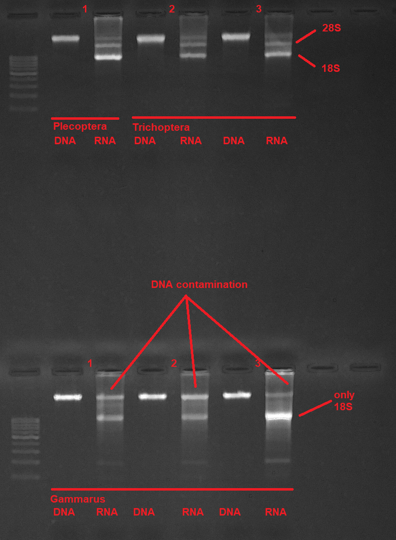
The described protocol was tested with different kinds of invertebrate samples, we expect it to work with all animal tissue. Top row: Plecoptera sample and two Trichoptera samples.Lower row: Three Gammarus samples.28S/18S bands are clearly visible and should have a clear band. Genomic DNA is free from RNA contamination. There is some DNA contamination in the RNA extracts. If DNA-free RNA is needed for downstream analysis consider treating the RNA samples with DNase and cleaning them up with an RNA cleanup protocol afterward (see RNA Cleanup with magnetic beads).

推荐阅读

Nature Protocols
Protocols IO
Current Protocols
扫码咨询